


Contents
Uterus
The uterus (from Latin uterus), is a hollow organ belonging to the female reproductive system and intended to accommodate and promote the development of the fertilized egg.
Anatomy of the uterus
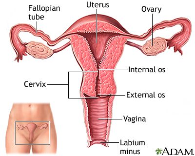
Location. The uterus is located in the pelvis at the back of the bladder, and in front of the rectum. The uterus is in the form of an inverted pyramid. In its upper part, two uterine tubes, or fallopian tubes, are inserted on each side face. Its lower part opens onto the vagina. (1)
Structure. The uterus is a hollow organ with thick walls, especially muscular ones. It is made up of two parts (1) (2):
- The body of the uterus is the largest part. It is located from the bottom of the uterus, the upper rounded part where the fallopian tubes are inserted, until the narrowing making the junction between the body and the cervix, called the isthmus of the uterus.
- The cervix is the narrowed part made up of two parts:
– The endocervix, or endocervical canal is the internal part of the cervix which starts from the isthmus and continues up to the opening opening into the vagina.
– The exocervix, corresponds to the external part of the cervix and is located in the upper part of the vagina.
Wall. The wall of the uterus is made up of three layers (3):
- The perimetrium which corresponds to the outer layer enveloping the body and part of the cervix.
- The myometrium which constitutes the middle layer made up of smooth muscles
- The endometrium which constitutes the inner layer lining the uterus and having glandular cells.
Support. Different ligaments support the uterus, in particular the uterosacral ligaments, or the round ligaments of the uterus. (1)
Uterine physiology
Role during pregnancy. The uterus is primarily intended to accommodate the embryo. During fertilization of the egg, the latter will implant itself in the endometrium at the level of the body of the uterus.
Menstrual cycle. It constitutes the set of modifications of the female genital apparatus in order to be able to receive a fertilized egg. In the absence of fertilization, the endometrium, the lining of the uterine body, is destroyed and evacuated through the cervix and then through the vagina. This phenomenon corresponds to menstrual periods.
Pathologies of the uterus
Cervical dysplasia. Dysplasias are precancerous lesions. They most often develop at the junction area between the cervix and the body of the uterus. They can extend to either side of the ectocervix and endocervix.
Human papillomavirus. Human papillomavirus (HPV) is a sexually transmitted virus. It comes in different forms: some can cause benign lesions in the cervix while others contribute to the development of precancerous lesions. In the latter case, the human papillomavirus is said to be potentially oncogenic or “at high risk” (4).
Benign tumors. Benign (non-cancerous) tumors can develop (3).
- Uterine fibroids. This benign tumor develops from muscle cells, mainly those of the muscular wall of the uterus.
- Endometriosis. This pathology corresponds to the development of endometrial tissue outside the uterus.
Uterus cancer. Different types of cancer can develop in the uterus.
- Endometrial cancer. This cancer develops in the endometrial cells of the uterine body. It represents the majority of uterine cancer cases.
- Cervical cancer Cervical cancer can occur when precancerous lesions, including cervical dysplasia, develop into cancer cells.
Treatments for the uterus
Surgical treatment. Depending on the pathology and its progress, a surgical intervention may be performed such as the removal of part of the uterus (conization).
Chemotherapy, radiotherapy, targeted therapy. Cancer treatment can take the form of chemotherapy, radiotherapy or even targeted treatment.
Uterine examinations
Physical examination. First, a physical examination is performed to assess the symptoms and characteristics of the pain.
Medical imaging exam Pelvic ultrasound, CT scan, or MRI can be used to confirm a diagnosis in the uterus.
Hysterography. This examination allows the observation of the uterine cavity.
Colposcopy: This test allows you to observe the walls of the cervix.5
Biopsy: Performed under colposcopy, it consists of a tissue sample.
Pap smear: This consists of a sample of cells from the upper level of the vagina, ectocervix and endocervix.
HPV screening test. This test is performed to screen for human papillomavirus.
History and symbolism of the uterus
Since 2006, a vaccine has been available for the prevention of infections due to human papillomavirus. This medical progress was made possible thanks to the work of the virologist Harald zur Hausen, Nobel Prize winner in medicine in 20086 After more than 10 years of research, he has succeeded in demonstrating the relationship between infections caused by a human papillomavirus and the occurrence of Cancer.