


Contents
Ureter
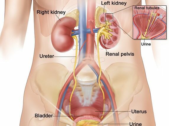
The ureter (from the Greek urêtêr) is a conduit in the urinary tract carrying urine from the kidneys to the bladder.
Anatomy of the ureters
Position. There are two ureters. Each ureter starts from the pelvis, part of the kidney accumulating urine, descends along the lumbar region before ending their journey by inserting through the wall of the postero-inferior surface of the bladder (1).
Structure. The ureter is a duct measuring between 25 and 30 cm long, with a diameter ranging from 1 to 10 mm, and presenting three areas of strictures (2). Muscular and elastic, its wall is made up of three layers (3):
- The detrusor which is the outer layer made up of smooth muscle tissue
- The lamina propria which is the intermediate layer of connective tissue comprising in particular nerves and blood vessels.
- The urothelium which is the inner layer of mucous membrane made up of urothelial cells.
Function of the ureter
Excretion of metabolic waste. The function of the ureters is to excrete the waste products concentrated in the urine, transporting it from the pelvis of the kidneys to the bladder before its elimination (2).
Pathologies and diseases of the ureters
Urinary lithiasis. This pathology corresponds to the formation of stones, concretions formed of mineral salts, at the level of the ureters. These calculations will lead to an obstruction of the ducts. This pathology can be manifested by severe pain called renal colic. (4)
Ureter malformations. There are many developmental abnormalities that can affect the ureter. For example, the defect in vesico-uteric reflux is caused by too short a segment of the ureter at the level of the bladder, which can cause infections (5).
Ureteral cancer. The cells of the ureter can be affected by benign (non-cancerous) tumors or malignant (cancerous) tumors. The latter are mainly linked to urothelial carcinoma, the cancer cells of which originate from the urothelium (3). This type of cancer is also very present in cases of bladder cancer.
Ureter treatments
Medical treatment. Depending on the pathology diagnosed, different drugs may be prescribed such as antibiotics or painkillers.
Surgical treatment. Depending on the pathology diagnosed, surgery may be performed. In the case of ureteric cancer, different operations may be performed depending on the stage and evolution of the tumor: removal of the tumor by endoscopic surgery, partial ablation by segmental resection or ablation total ureter by radical nephro-ureterectomy (3).
Chemotherapy, radiotherapy. Depending on the stage of the tumor, chemotherapy or radiotherapy sessions may be set up. (6)
Ureter examinations
Urine cytobacteriological examination (ECBU). In case of ureteric infections, this test can be done to identify bacteria present in the urine and their sensitivity to antibiotics. This examination is carried out in particular in the event of complicated cystitis.
Medical imaging exams. Different medical imaging exams can be used to analyze the bladder: ultrasound, intravenous urography, retrograde cystography or uroscanner.
Ureteroscopy.This endoscopic examination is performed to analyze the walls of the ureters. It is practiced in particular to treat urinary stones in the event of urinary lithics.
Urinary cytology. This test can identify the presence of cancer cells in the urine.
History and symbolism of the ureter
Dating from ancient Egypt and practiced until the 7th century, uroscopy is a pioneering medical practice in urology. Replaced today by urine strips, uroscopy consisted of the visual examination of urine in order to identify the development of certain pathologies (XNUMX).