


Retinal detachment: causes, symptoms, treatment
The retina, a membrane essential to our vision, can in rare situations become detached. This is a serious problem, to be detected as early as possible to limit the consequences.
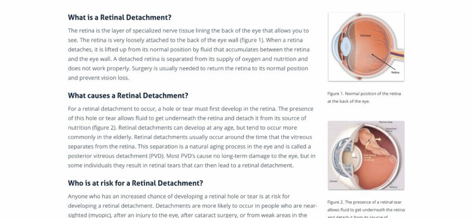
Lurking at the back of our eye, the retina is a membrane studded with nervous tissue and connected to the optic nerve. It is on it that the photons of light rays are received, before being transmitted to the brain. However, this membrane is not that strong. It relies on two others to form a complete eye. It therefore happens that the retina takes off, partially or totally, which can lead to a blindness total.

How does the retina peel off?
The human eyeball is made up of three successive layers of membranes, called tunics. The first, the fibrous tunic is the one we can see: white, it covers the eye up to the cornea in the front. The second, located just below, is the uveal tunic (or uvée). It is made up at the front of the iris, and at the back of a layer called the choroid. Finally, glued to the uveal tunic, we find the famous nervous tunic, the retina.
The retina itself breaks down into different layers. Thus, when we speak of detachment of the retina, it is above all that of the neural retina compared topigment epithelium, its outer wall. Their connection is indeed very fragile, and shocks or lesions can lead to the creation of openings, within which a liquid such as vitreous can enter, and accelerate the detachment process.
The causes of retinal detachment
Retinal detachment affects only one in 10 people on average, but can have various causes related to the patient profile.
Myopia
Nearsighted people have a deeper-than-average eye, which is why their focal point is “in front” of the retina. The latter therefore turns out to be thinner than average, and therefore has a greater risk of peeling off or tearing one day. Myopic people therefore have every interest in seeing their ophthalmologist regularly in order to quickly detect the first signs.
Genetic
Like nearsightedness, genetics sometimes work against us and cause eye deformities. Retina fragile, too thin, or poorly fixed, it happens that the cause is genetic in rare cases. It will then be necessary to be careful to know whether the operation will be effective or not.
Glaucoma and cataracts
People with glaucoma or having had a cataract operation are also more likely to have their retina detachment. Blame it on an already weakened eye,
Athletes: martial arts and boxing
Our eye is not that strong, and the absence of pain when impacting it too often prevents us from realizing it. Thus, retinal detachment is more common in athletes who regularly take blows to the face. By being mistreated in this way, the eye moves in its orbit, bumps into itself, and the whole is weakened, including the retina.
Symptoms of detachment
It will be much easier to treat a retinal detachment if it is just incipient, compared to the more advanced one. The most severe cases will require much more complex surgeries. So here is a list of warning signs. Some are not necessarily the sign of a detachment, others yes. In all cases, it is better to seek the advice of an ophthalmologist quickly if at least two of these symptoms are present:
Mouches floaters
It is the most common symptom of a detachment, when the sight is filled with “flying flies”, that is to say small black dots, always present. However, this is not necessarily a sign that the retina is peeling off, and it should not be confused with the flying bodies within the vitreous, a more temporary gene.
Eclairs
If one or more lightning flashes appear in your field of vision (outside the storm period!), It may be that the retina has suddenly detached in one or more areas. By being suddenly disconnected from the nervous system, the impulse received by the brain resembles that of a lightning bolt.
Dark spots, obscured vision
If the retina is peeling off, then some areas of your field of vision will be dark. If these areas are on the periphery of the retina, then it may be more difficult to spot it quickly. It is therefore ideal to perform an examination while at rest, if you have any of the other symptoms (fly flies or lightning), to see if you have not noticed a dark area. In the worst case, if the retina has detached at its most sensitive point, the macula, it is your central vision that disappears. In this case, you must consult the ophthalmologist emergency room very quickly.
Complete blindness
It is the most abrupt symptom, but if the retinal detachment is not treated quickly, it can completely detach from the optic nerve, and the eye simply cannot see anything.
Treatments
The treatment of retinal detachment is complex, and depends on its severity.
Surgery laser
For mild detachments, laser surgery can reattach the retina and cauterize the tearing areas.
Vitrectomie
For the most serious cases, the practitioner must be able to manually repair the retina. In order to access the back of the eye, the doctor will therefore have to remove the vitreous, gelatinous liquid within the eyeball. To do this, he pierces openings in the side of the eye, sucks in the vitreous, and can then manually reattach the retina. The vitreous body is then replaced with a gel or silicone oil.
Strapping
The strapping consists of encircling the eye, in order to press on both sides to reattach the retina, if it is not yet very detached.
Cryo-indentation
Applying very cold gas to the eye can cause scarring of the retina in the area that is peeling or tearing. This technique avoids entering the eye but is reserved for light detachments. Sometimes, however, it will be necessary to place ties within the eye in order to fix the retina for good while it repairs itself.