Contents
Douglas cul-de-sac: role, anatomy, effusion
What is Douglas’s cul-de-sac?
Douglas is the name of a Scottish anatomist doctor James Douglas (1675-1742), who gave his name to the various cul-de-sac terms of Douglas and to the pathologies linked to it: douglassectomy, douglascele, douglassite, Douglas line , etc.
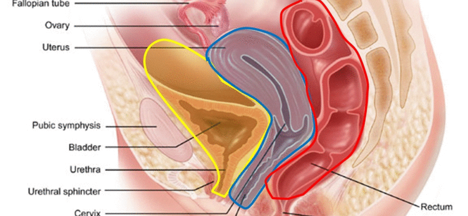
The Douglas’ cul-de-sac is described by anatomists as a fold of the peritoneum located between the rectum and the uterus, forming a cul-de-sac.
Location of the Douglas cul-de-sac
Douglas’ cul-de-sac is located at a distance below the umbilicus of 4 to 6 cm. It is the lowest point of the peritoneal cavity, which is itself formed by the peritoneum, a serous membrane that lines the abdominal cavity.
At men’s
In men, this cul-de-sac is located between the bladder and the rectum. It is simply the lower end of the peritoneal cavity, between the posterior surface of the bladder and the anterior surface of the rectum.
In women
For women, the Douglas pouch is also called the recto-uterine pouch, it is located between the rectum and the uterus. It is therefore limited behind by the rectum, in front by the uterus and the vagina; and laterally by the recto-uterine folds.
Role of Douglas’ cul-de-sac
Its role is to support the organs and protect them against infections.
Operation
It is made up of dense connective tissue containing collagen-like proteins, and elastic fibers. This solid membrane is also called aponeurosis.
This membrane has the capacity to secrete serosities, a sort of lymphatic fluid equivalent to the liquid part of the blood called plasma.
Serum forms in the serous membranes which are the membranes that line the closed cavities of the body.
Douglas Cul-de-Sac Examinations
Douglas’ cul-de-sac is accessible by a vaginal examination in women, by a rectal examination in men.
This digital palpation examination is normally painless.
If this touch causes pain, the patient cries out because the pain is so violent. This cry is known by health professionals to be “the cry of the Douglas” as the symptoms are so specific.
Associated diseases and treatments of Douglas’ cul-de-sac
Palpation shows an intraperitoneal effusion, abscess or solid tumor. In case of abscess, palpation can be very painful.
This pain can be the sign of a multitude of pathologies which can range from an ectopic pregnancy in women, a hernia or even a douglassitis.
Ectopic (or ectopic) pregnancy
An ectopic (or ectopic) pregnancy develops outside the uterine cavity:
- in a fallopian tube, it is a tubal pregnancy;
- in the ovary, it is an ovarian pregnancy;
- in the peritoneal cavity, it is an abdominal pregnancy.
In the case of an ectopic pregnancy, the vaginal examination of the obstetrician or midwife is extremely painful (Douglas pain) and may be accompanied by syncope, pallor, acceleration pulse, fever, bloating. The Douglas can be filled with sepia brown colored blood.
The effusion of the small pelvis, therefore behind this vaginal cul-de-sac, behind the uterus, is frequently encountered in the event of a ruptured ectopic pregnancy. This rupture causes a blood effusion which accumulates behind this cul-de-sac. Its palpation is then extremely painful and quite significant for the diagnosis.
Elytrocellular or double glazed
This organ descent (or prolapse) is caused by a hernia of the intestine that has descended into the Douglas’ cul-de-sac and which pushes the posterior vaginal wall back through the vulva.
Douglassite
Douglassitis is a chronic inflammation of the peritoneum which is located in the Douglas-fir sac. It is usually caused by an intraperitoneal effusion (in the peritoneum, a tumor, a collection of blood from a hemorrhage caused by a GEU (ectopic pregnancy) or an abscess or abscess.
The doctor performs a rectal (for the man) or vaginal (for the woman) to know the state of the cul-de-sac.
The different interventions
When the effusion needs to be removed, the doctor performs drainage. For women, it is a colpotomy, an intervention through the vaginal wall and for men this intervention is called a rectotomy, because the intervention is done through the rectal wall.
Douglas Cul-de-Sac Treatments
When Douglas’ cul-de-sac is filled with blood or fluid, it is therefore necessary to perform drainage, particularly in women through the vaginal walls. This gesture is the colpotomy.
In humans, drainage is sometimes necessary as well. In this case it is necessary to perform it through the anterior wall of the rectum, this intervention is called a rectotomy.
The localization of an effusion can be confirmed by an ultrasound and a puncture precise its nature.
Douglassectomy
Douglassectomy is a surgical procedure that involves removing Douglas’ cul-de-sac. It is performed by laparoscopy or through an opening in the abdomen called a laparotomy.


