


Contents
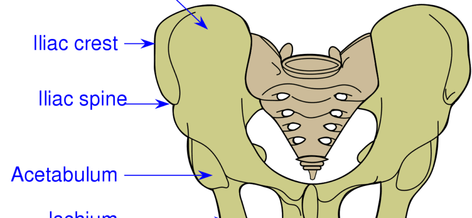
Iliac crest
The iliac crest forms part of the ilium or ilium, bone making up the upper part of the coxal bone, or iliac bone.
Pelvic anatomy
Position. The iliac crest is the top of the hip bone, or iliac bone. Located at the level of the pelvic girdle (1), the latter is an even bone made up of three bones welded together (2):
- The ilium which constitutes the upper part of the coxal bone.
- The pubis which designates the antero-inferior part.
- The ischium which corresponds to the postero-inferior part.
Structure. The iliac crest forms the thickest upper edge of the ilium. The latter is a large, flared bone constituting the largest portion of the hip bone. It is made up of two parts (1) (2):
- The body of the ilium on its lower part.
- The wing of the ilium, wing-shaped, on its upper part.
The iliac crest begins at the level of the anterosuperior iliac spine, the bony protrusion constituting the forward end and ends at the level of the postero-superior iliac spine, the bony protrusion constituting the posterior end (1) ( 3).
Muscle insertion. The iliac crest serves as the insertion zone for many muscles (4). In the front, we can distinguish the transverse muscle of the abdomen, as well as the internal and external oblique muscles of the abdomen. At the back, we find the square muscle of the lumbar muscles and the latissimus dorsi muscle.
Physiology / Histology
Muscle insertion zone. The iliac crest serves as an attachment area for various muscles in the abdomen.
Fractures. The ilium, including the iliac crest, can fracture, including pain in the hip.
Bone diseases. Certain bone pathologies can affect the ilium, such as osteoporosis, which is a loss of bone density and is generally found in people over the age of 60 (5).
Tendinopathies. They designate all the pathologies that can occur in the tendons, in particular those associated with the muscles attached to the iliac crest. The causes of these pathologies can be varied. The origin can be intrinsic as well with genetic predispositions, as extrinsic, with for example bad positions during the practice of sport.
- Tendinitis: It is an inflammation of the tendons.
Treatments
Medical treatment. Depending on the pathology diagnosed, certain drugs may be prescribed to reduce pain.
Orthopedic treatment. Depending on the type of fracture, the installation of a plaster or a resin can be carried out.
Surgical treatment. Depending on the pathology and its evolution, a surgical intervention may be implemented.
Physical treatment. Physical therapy, through specific exercise programs, can be prescribed such as physiotherapy or physiotherapy.
Iliac crest examination
Physical examination. First, a clinical examination is performed to identify painful movements.
Medical imaging exam. Depending on the suspected or proven pathology, additional examinations can be performed such as an X-ray, an ultrasound, a CT scan, an MRI, a scintigraphy or even a bone densitometry.
Medical analysis. In order to identify certain pathologies, blood or urine analyzes can be carried out such as, for example, the dosage of phosphorus or calcium.
Anecdote
Work on the human skeleton has revealed a change in the size and shape of the pelvic bones during evolution. It seems that the transition from flat bones to curved bones, as well as a longer growth allowed the acquisition of bipedalism. The lower limbs thus became closer and closer together and would have allowed locomotion as well as walking (6).