


Contents
Diaphragm
The diaphragm is the essential muscle in the mechanics of breathing.
Anatomy of the diaphragm
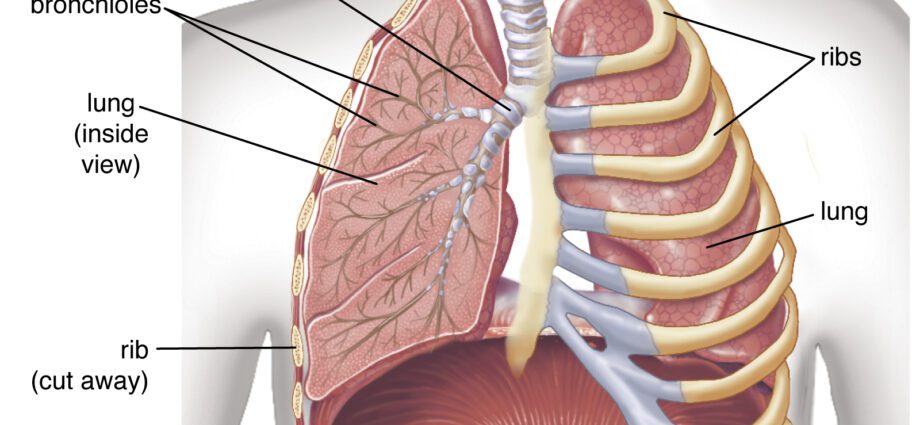
The diaphragm is an inspiratory muscle located under the lungs. It separates the chest cavity from the abdominal cavity. In the shape of a dome, it is marked with two domes on the right and on the left. They are asymmetrical, the right diaphragmatic dome is normally 1 to 2 cm higher than the left dome.
The diaphragm is made up of a central tendon, the tendon center of the diaphragm or phrenic center. At the periphery, muscle fibers connect at the level of the sternum, ribs and vertebrae.
It has natural orifices which allow the passage of organs or vessels from one cavity to another. This is the case, for example, with the esophageal, aortic or inferior vena cava orifices. It is innervated by the phrenic nerve which causes it to contract.
Physiology of the diaphragm
The diaphragm is the main respiratory muscle. Associated with the intercostal muscles, it ensures the mechanics of breathing by alternating the movements of inspiration and expiration.
On inspiration, the diaphragm and intercostal muscles contract. As it contracts, the diaphragm lowers and flattens. Under the action of the intercostal muscles, the ribs go up which raises the rib cage and pushes the sternum forward. The thorax then increases in size, its internal pressure decreases which causes a call for outside air. Result: air enters the lungs.
The frequency of the diaphragm contraction defines the respiratory rate.
On exhalation, the diaphragm and intercostal muscles relax, causing the ribs to descend as the diaphragm rises back to its original position. Gradually, the rib cage lowers, its volume decreases which increases its internal pressure. As a result, the lungs retract and air escapes from them.
Diaphragm pathologies
Hiccups : designates a succession of involuntary and repeated spasmodic contractions of the diaphragm associated with a closure of the glottis and often a contraction of the intercostal muscles. This reflex occurs suddenly and uncontrollably. It results in a series of characteristic sonic “hics”. We can distinguish the so-called benign hiccups which last no more than a few seconds or minutes, and the chronic hiccups, much rarer, which can last from several hours to several days and which generally affects people over 50 years old.
Post-traumatic ruptures : diaphragm ruptures which take place following trauma to the thorax, or wounds by bullets or bladed weapons. The rupture usually occurs at the level of the left dome, the right dome being partially hidden by the liver.
Transdiaphragmatic hernia : rise of an organ in the abdomen (stomach, liver, intestines) through an orifice in the diaphragm. The hernia can be congenital, the hole through which the migrating organ passes is a malformation present from birth. It can also be acquired, the hole is then the consequence of an impact during a road accident for example; in this case we speak of diaphragmatic eventration. It is a rare condition that affects nearly one in 4000 babies.
Elevation of a diaphragmatic dome : the right dome is normally 1 to 2 cm higher than the left dome. There is an “elevation of the right dome” when the distance exceeds 2 cm from the left dome. This distance is checked on a chest X-ray taken in deep inspiration. We speak of “elevation of the left dome” if it is higher than the right or simply at the same level. It may reflect an extra-diaphragmatic pathology (ventilation disorders or pulmonary embolism for example) or a diaphragmatic pathology (traumatic lesions of the phrenic nerve or hemiplegia for example) (5).
Tumors : they are very rare. Most often these are benign tumors (lipomas, angio and neurofibromas, fibrocytomas). In malignant tumors (sarcomas and fibrosarcomas), there is often a complication with pleural effusion.
Neurological pathologies : Any damage to a structure located between the brain and the diaphragm can have consequences on its functioning (6).
For example, Guillain-Barré syndrome (7) is an inflammatory autoimmune disease that attacks the peripheral nervous system, in other words the nerves. It manifests itself by muscle weakness which can go as far as paralysis. In the case of the diaphragm, the phrenic nerve is affected and breathing disturbances appear. Under treatment, the majority of affected people (75%) recover their physical capacities.
Amyotrophic lateral sclerosis, or Charcot’s disease, is a neurodegenerative disease characterized by progressive muscle paralysis due to the degeneration of motor neurons that send orders for movement to muscles. As the disease progresses, it can affect the muscles necessary for breathing. After 3 to 5 years, Charcot’s disease can therefore cause respiratory failure which can lead to death.
Case of hiccups
Only hiccups can be the subject of a few measures. It is difficult to prevent its appearance which is quite random, but we can try to reduce the risks by avoiding eating too quickly, as well as excess tobacco, alcohol or carbonated drinks, stressful situations or sudden changes in temperature.
Diaphragm examinations
The diaphragm is difficult to study on imaging (8). Ultrasound, CT and / or MRI are often in addition to standard radiography to confirm and refine the diagnosis of a pathology.
Radiography: a medical imaging technique that uses X-rays. This examination is painless. The diaphragm is not directly visible on a chest x-ray, but its position can be identified by the line that marks the lung-liver interface on the right, lung-stomach-spleen on the left (5).
Ultrasound: a medical imaging technique based on the use of ultrasound, inaudible sound waves, which make it possible to “visualize” the interior of the body.
MRI (magnetic resonance imaging): medical examination for diagnostic purposes carried out using a large cylindrical device in which a magnetic field and radio waves are produced to generate very precise images, in 2D or 3D, of parts of the body or organs internal (here the diaphragm).
Scanner: diagnostic imaging technique which consists of creating cross-sectional images of a certain part of the body, using an X-ray beam. The term “scanner” is in fact the name of the device, but we commonly used to refer to the exam (computed tomography or CT scan).
Anecdote
In human anatomy, the word diaphragm is also used to refer to the iris of the eye. The iris controls the amount of light that enters the eye. This function is worth it to be compared to the diaphragm of a camera.