


Contents
Small intestine
The small intestine (from the Latin intestinum, from intestine, meaning “inside”) is an organ of the digestive tract.
Anatomy of the small intestine
Localisationot. 5 to 7 meters long and 3 cm in diameter, the small intestine follows the stomach and is extended by the large intestine (1).
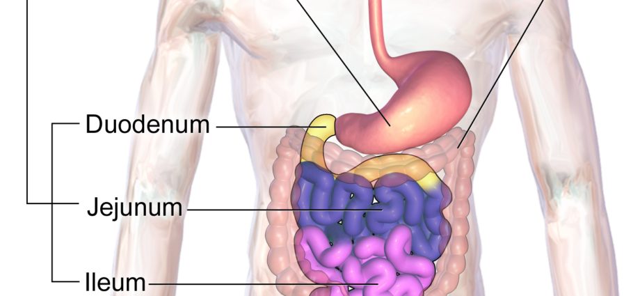
Structure. The small intestine is made up of three segments (1) (2):
- The duodenum is located between the pylorus of the stomach and the duodeno-jejunal angle. C-shaped and deeply located, it forms the fixed part of the small intestine. The excretory ducts from the pancreas and the bile duct arrive at this segment.
- The jejunum starts at the duodeno-jejunal angle and extends to the ileum. It constitutes, with the ileum, the major part of the small intestine.
- The ileum follows the jejunum and extends to the ileocecal valve, leading to the large intestine. The ileum and jejunum constitute the mobile part of the small intestine.
Wall. The small intestine is made up of 4 envelopes (1):
- The mucous membrane is the inner layer containing many glands, secreting in particular a protective mucus.
- The submucosa is the intermediate layer made up in particular of vessels and nerves.
- The muscularis is the outer layer made up of muscle fibers.
- The serous membrane, or peritoneum, is an envelope lining the outer wall of the small intestine.
Physiology / Histology
Digestion. Digestion takes place mainly in the small intestine, and more particularly in the duodenum through digestive enzymes and bile acids. Digestive enzymes originate from the pancreas through the excretory ducts, while bile acids originate from the liver through the bile ducts (3). Digestive enzymes and bile acids will transform chyme, a liquid comprising food predigested by digestive juices from the stomach, into chyle, a clear liquid containing dietary fibers, complex carbohydrates, simple molecules, as well as nutrients (4).
Absorption. For its activity, the body will absorb certain elements such as carbohydrates, fats, proteins, electrolytes, vitamins, as well as water (5). The absorption of the products of digestion takes place mainly in the small intestine, and mainly in the duodenum and jejunum.
Protection of the small intestine. The small intestine defends itself against chemical and mechanical attacks by secreting mucus, protecting the mucous membrane (3). The small intestine is also protected from contamination by bacteria in the large intestine thanks to the ileocecal valve.
Pathology and disease of the small intestine
Chronic inflammatory bowel disease. These diseases correspond to inflammation of the lining of part of the digestive system, such as Crohn’s disease. Symptoms include severe abdominal pain and diarrhea (6).
Irritable bowel syndrome. This syndrome is manifested by hypersensitivity of the wall of the intestine and irregularity in muscle contractions. It manifests itself through various symptoms such as diarrhea, constipation, or abdominal pain. The cause of this syndrome is still unknown today.
Bowel obstruction. It indicates a stop of the functioning of the transit, causing intense pain and vomiting. Intestinal obstruction can be of mechanical origin with the presence of an obstacle during transit (gallstones, tumors, etc.) but can also be chemical by being linked to an infection of a nearby tissue, for example during peritonitis.
Peptic ulcer. This pathology corresponds to the formation of a deep wound in the wall of the stomach or that of the duodenum. Peptic ulcer disease is often caused by bacterial growth but can also occur when taking certain medications (7).
Treatments
Medical treatment. Depending on the pathology diagnosed, certain drugs may be prescribed such as anti-inflammatory drugs or analgesics.
Surgical treatment. Depending on the pathology and its evolution, a surgical intervention may be implemented.
Examination of the small intestine
Physical examination. The onset of pain begins with a physical examination to assess symptoms and identify the causes of the pain.
Biological examination. Blood and stool tests may be done to make or confirm a diagnosis.
Medical imaging exam. Depending on the suspected or proven pathology, additional examinations may be performed such as an ultrasound, a CT scan or an MRI.
Endoscopic examination. An endoscopy can be done to study the walls of the small intestine.
History
In 2010, researchers from Inserm in Nantes published their research results on the effects of Parkinson’s disease on digestive neurons in the scientific journal Plos One. They have shown that the lesions of Parkinson’s disease affect not only the cells of the central nervous system, but also those of the enteric nervous system, and more precisely that of the digestive system. This discovery could allow an early diagnosis of Parkinson’s disease (8).