


Contents
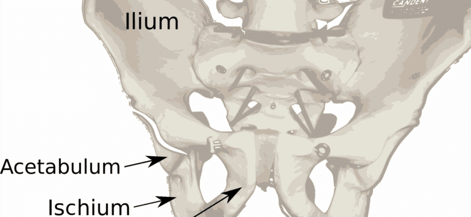
Ilium
The ilium (from the lower Latin ilium, coming from the classical Latin ilia, meaning flanks), also called ilium, is a bone constituting the upper part of the coxal bone, or iliac bone, located at the level of the pelvic girdle (1) .
Anatomy of the ilium
Position. The ilium is the upper part of the hip bone, or iliac bone. The latter is an even bone made up of three bones fused together: the ilium, the pubis which designates the antero-inferior part and the ischium which constitutes the postero-inferior part (2).
Structure. The ilium is a large, flared bone making up the largest portion of the hip bone. It is made up of two parts (1) (2):
- The body of the ilium, which includes on its lower part a part of the acetabulum, a cavity involved in the coxofemoral joint;
- The wing of the ilium, wing-shaped, which forms the upper part of the ilium.
Insertions and passages. The ilium has different attachment points (1) (3):
- The iliac crest forms the thicker upper border. It serves as an insertion zone for many muscles. Called the upper iliac spines, the upper ends of the iliac crest serve as attachment points for the muscles of the trunk, hips, and thighs. We distinguish :
– The antero-superior iliac spine, bony projection, which constitutes the front end of the iliac crest.
– The posterior superior iliac spine, bony projection, which constitutes the rear end of the iliac crest.
- The large ischial notch, located under the iliac spines, serves as a passage for the sciatic nerve.
- The gluteal lines, located on the posterior and lateral surface, serve as points of attachment to the gluteal muscles.
- The atrial surface of the ilium, located at the back of the ilium wing, articulates with the sacrum to form the sacroiliac joint.
Physiology / Histology
Weight transmission. The ilium and its articulation with the sacrum are involved in the transmission of weight from the upper part of the body to the neck of the femur and then to the lower limbs (3).
Muscle insertions zone. The ilium serves as an attachment area for many muscles.
Sciatic nerve passage area. The ilium allows passage from the sciatic nerve to the thigh.
Pathologies associated with ilium
fractures. The ilium can suffer from fractures such as fracture of the acetabulum. These fractures are manifested in particular by pain in the hip.
Bone diseases. Certain bone pathologies can affect the ilium, such as osteoporosis, which is a loss of bone density and is generally found in people over the age of 60 (4).
Ankylosing spondylitis. This rheumatic inflammatory disease can affect the joints of the vertebrae, and more particularly the sacroiliac joints. It causes pain as well as stiffness in the pelvis.
Treatments
Medical treatment. Depending on the pathology diagnosed, certain drugs may be prescribed to reduce pain.
Orthopedic treatment. Depending on the type of fracture, the installation of a plaster or a resin can be carried out.
Surgical treatment. Depending on the pathology and its evolution, a surgical intervention may be implemented.
Physical treatment. Physical therapy, through specific exercise programs, can be prescribed such as physiotherapy or physiotherapy.
L’ilion exam
Physical examination. First, a clinical examination is performed to identify painful movements.
Medical imaging exam. Depending on the suspected or proven pathology, additional examinations can be performed such as an X-ray, an ultrasound, a CT scan, an MRI, a scintigraphy or even a bone densitometry.
Medical analysis. In order to identify certain pathologies, blood or urine analyzes can be carried out such as, for example, the dosage of phosphorus or calcium.
History
Work on the human skeleton has revealed a change in the size and shape of the pelvic bones during evolution. It seems that the transition from flat bones to curved bones, as well as a longer growth allowed the acquisition of bipedalism. The lower limbs thus became closer and closer together and would have allowed locomotion as well as walking (5).