


Contents
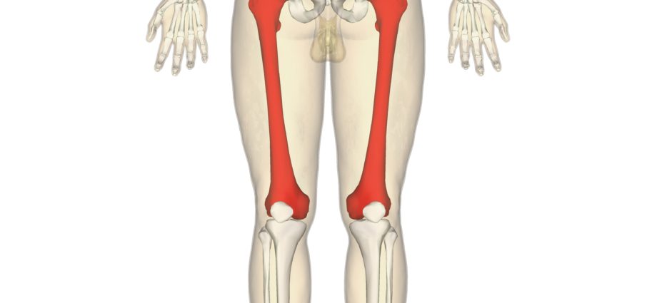
Femur
The femur (from Latin femur) is the only thigh bone located between the hip and the knee.
Anatomy of the femur
Overall structure. Elongated in shape, the femur is the longest bone and represents on average a quarter of the body’s size. (1) It is also the largest bone in the human body and is made up of three parts:
- a proximal end, located at the hip;
- a distal end, located at the knee
- a diaphysis, or body, central part of the bone located between the two ends.
Joints. The proximal end of the femur is made up of three parts (1):
- the head of the femur, located in the acetabulum, the articular cavity of the hip bone, which forms the hip;
- the neck of the femur which connects the head to the shaft;
- two trochanters, bony protrusions, which are positioned at the level of the connection of the neck and the head.
The distal end of the femur consists of:
- femoral condyles, or articular surfaces, which articulate with the condyles of the tibia to form the knee;
- a patellar surface that articulates with the patella;
- epicondyles, bony protrusions, and tubercles, which serve as points of attachment to muscles and ligaments. (1)
Functions of the femur
Weight transmission. The femur transmits the body’s weight from the hip bone to the tibia. (2)
Body dynamics. The joints of the femur at the hip and knee are involved in the body’s ability to move and maintain the upright posture. (2)
Femur pathologies
Fractures of the femur. The most common femoral fractures are those in the neck of the femur, especially in older people with osteoporosis. They can also occur between the trochanters, at the proximal end, and at the shaft (1). Fractures are manifested by pain in the hip.
Femoral head epiphysis. Epiphysiolysis is manifested by an abnormality of the epiphyseal plaque, which refers to the plaque at the end of a long bone such as the femur. This pathology can develop at the proximal end of the femur causing the head of the femur to detach from the neck of the femur. This detachment can also cause other abnormalities such as a coxa vara, deformation of the upper part of the femur. (1)
Thigh stick, thigh valga. These problems correspond to a deformation of the upper part of the femur by modification of the angle of inclination between the neck and the body of the femur. This angle is normally between 115 ° and 140 °. When this angle is abnormally lower, we speak of stick thigh, while when it is abnormally higher, it is a thigh valgus. (1)
Bone diseases.
- Osteoporosis. This pathology constitutes a loss of bone density which is generally found in people over the age of 60. It accentuates bone fragility and promotes bills. (3)
- Bone cancer. Metastases can develop in the bones. These cancer cells usually originate from primary cancer in another organ. (4)
- Bone dystrophy. This pathology constitutes an abnormal development or remodeling of bone tissue and includes many diseases. One of the most common, Paget’s disease (5) causes bone densification and deformation, leading to pain. Algodystrophy refers to the appearance of pain and / or stiffness following a trauma (fracture, surgery, etc.).
Femur treatments
Medical treatment. Depending on the disease diagnosed, different treatments may be prescribed to regulate or strengthen bone tissue, as well as to reduce pain and inflammation.
Surgical treatment. Depending on the type of fracture, a surgical operation can be performed with the placement of pins, a screw plate, an external fixator or in some cases a prosthesis.
Orthopedic treatment. Depending on the type of fracture, the installation of a plaster or a resin can be carried out.
Physical treatment. Physical therapies, such as physiotherapy or physiotherapy, may be prescribed.
Hormonal treatment, radiotherapy or chemotherapy. These treatments may be prescribed depending on the stage of cancer progression.
Femur examinations
Physical examination. Diagnosis begins with an assessment of lower limb pain to identify its causes.
Medical imaging examination. Depending on the suspected or proven pathology, additional examinations may be performed such as an X-ray, an ultrasound, a CT scan, an MRI, a scintigraphy or even a bone densitometry.
Medical analysis. In order to identify certain pathologies, blood or urine analyzes can be carried out such as, for example, the dosage of phosphorus or calcium.
Bone biopsy. In some cases, a bone sample is taken to confirm a diagnosis.
History and symbolism of the femur
In December 2015, the magazine PLOS ONE unveiled an article relating the discovery of a human femur from a premodern species. (6) Discovered in 1989 in China, this bone was not studied until 2012. Dating back 14 years, this bone seems to belong to a species approaching theHomo handy orHomo erectus. Primitive humans could thus have survived until the end of the last Ice Age, 10 years ago. This discovery could suggest the existence of a new evolutionary lineage (000).