


Dendrites: a major role in information processing?
The human nervous system, of intense complexity, is made up of approximately 100 billion neurons, also called nerve cells. Neurons in the brain can communicate through synapses that transmit the nerve signal from one neuron to another.
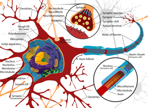
Dendrites are short, branched extensions of these neurons. Indeed, dendrites form the receptor part of the neuron: they are often represented as a sort of tree emerging from the neuronal cell body. In fact, the logical function of dendrites will therefore consist in collecting information at the level of the synapses which cover them, before routing them to the cell body of the neuron.
Anatomy of dendrites
Nerve cells are very different from other cells in the human body: on the one hand, their morphology is very particular and on the other hand, they operate electrically. The term dendrite comes from the Greek word Dendron, which means “tree”.
The three parts that make up the neuron
Dendrites are the main receptor parts of the neuron, also called a nerve cell. In fact, most neurons are made up of three main components:
- a cell body;
- two kinds of cellular extensions called dendrites;
- axons.
The cell body of neurons, also called soma, contains the nucleus as well as other organelles. The axon is a single, thin, cylindrical extension that directs the nerve impulse to another neuron or to other types of tissue. In fact, the only logical function of the axon is to drive, from one place in the brain to another, a message encoded in the form of a succession of action potentials.
What about dendrites more precisely?
A tree structure emerging from the cell body
These dendrites are short, tapered, and highly branched extensions, forming a sort of tree that emerges from the neuronal cell body.
The dendrites are indeed the receptor parts of the neuron: in fact, the plasma membrane of the dendrites contains multiple receptor sites for the binding of chemical messengers from other cells. The radius of the dendritic tree is estimated at one millimeter. Finally, many synaptic buttons are located on dendrites in places far from the cell body.
The ramifications of dendrites
Each dendrite emerges from the soma by a cone which extends into a cylindrical formation. Very quickly, it will then divide into two branch-daughter. Their diameter is smaller than that of the parent branch.
Then, each of the ramifications thus obtained divides, in turn, into two other, finer ones. These subdivisions continue: this is the reason why neurophysiologists metaphorically evoke “the dendritic tree of a neuron”.
Physiology of dendrites
The function of dendrites is to collect information at the level of the synapses (spaces between two neurons) which cover them. Then these dendrites will carry this information to the neuron’s cell body.
Neurons are sensitive to various stimuli, which they convert into electrical signals (called nervous action potentials), before in turn transmitting these action potentials to other neurons, muscle tissue or even to glands. And indeed, whereas in an axon, the electric impulse leaves the soma, in a dendrite, this electric impulse propagates towards the soma.
A scientific study made it possible, thanks to microscopic electrodes implanted in neurons, to evaluate the role that dendrites have in the transmission of nerve messages. It turns out that, far from being simply passive extensions, these structures play a major role in information processing.
According to this study published in Nature, the dendrites would therefore not only be simple membrane extensions involved in relaying the nerve impulse to the axon: they would in fact not be simple mediators, but they too would process information. A function that would increase the capacities of the brain.
So all the data seem to converge: dendrites are not passive, but are, in a way, minicomputers in the brain.
Anomalies / pathologies of dendrites
The abnormal functioning of the dendrites can be linked to dysfunctions relating to the neurotransmitters which excite them or, on the contrary, inhibit them.
The best known of these neurotransmitters are dopamine, serotonin or even GABA. These are dysfunctions of their secretion, which is too high or on the contrary too low, or even inhibited, which can be the cause of anomalies.
The pathologies caused by a failure in neurotransmitters are, in particular, psychiatric illnesses, such as depression, bipolar disorder or schizophrenia.
Psychic failures linked to poor regulation of neurotransmitters and therefore, downstream, to the functioning of dendrites, are now increasingly treatable. Most often, a beneficial effect on psychiatric pathologies will be obtained by an association between drug treatment and psychotherapeutic type monitoring.
Several types of psychotherapeutic currents exist: in fact, the patient can choose a professional with whom he feels confident, listened to and a method that suits him according to his past, his experience, and his needs.
There are in particular cognitive-behavioral therapies, interpersonal therapies or even psychotherapies more linked to a psychoanalytic current.
What diagnosis?
The diagnosis of a psychiatric illness, which therefore corresponds to a failure of the nervous system in which the dendrites play a crucial role, will be made by a psychiatrist. It will often take quite a long time to make a diagnosis.
Finally, it is important to know that the patient should not feel trapped in a “label” which would characterize him, but that he remains a full person, who will simply have to learn to manage his particularity. Professionals, psychiatrists and psychologists, will be able to help him in this direction.
History and symbolism
The date of introduction of the term “neuron” is set at 1891. This adventure, essentially anatomical at the outset, emerged in particular thanks to the black coloring of this cell, carried out by Camillo Golgi. But, this scientific epic, far from focusing only on the structural aspects of this discovery, gradually made it possible to conceive of the neuron as a cell being the seat of electrical mechanisms. It then appeared that these regulated reflexes, as well as complex brain activities.
It was mainly from the 1950s that many sophisticated biophysical instruments were applied to the study of the neuron, at the infra-cellular and then the molecular level. Thus, the electron microscopy made it possible to reveal the space of the synaptic cleft, as well as the exocytosis of neurotransmitter vesicles at the synapses. It was then possible to study the content of these vesicles.
Then, a technique called “patch-clamp” made it possible, from the 1980s, to study current variations through a single ion channel. We were then able to describe the intimate intracellular mechanisms of the neuron. Among them: the back-propagation of action potentials in dendrite trees.
Finally, for Jean-Gaël Barbara, neuroscientist and science historian, “gradually, the neuron becomes the object of new representations, like a special cell among others, while being unique by the complex functional meanings of its mechanisms«.
Scientists Golgi and Ramon y Cajal were awarded the Nobel Prize in 1906 for their work relating to the concept of neurons.