


Contents
Mesothelium, what is it?
The mesothelium is a membrane that lines most internal organs in order to cover and protect them. It is made up of two layers of flattened cells, one of which, the inner layer, envelops different organs such as the lungs, heart and stomach, and the second, the outer layer, forms a kind of sac surrounding the inner layer. . Fluid is present between these two layers of cells, which facilitates the movement of organs.
The mesothelium can sometimes be affected by benign tumors, and much more rarely, cancers called mesotheliomas. It is then in the pleura that it is most frequent, that is to say the mesothelium which covers the lung; in the vast majority of cases, it is due to exposure to asbestos. But this condition remains very rare, there are, according to figures from the High Authority for Health, 600 to 900 new cases identified each year in France.
Anatomy of the mesothelium
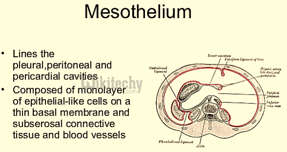
The mesothelium is made up of two layers of flattened cells called mesothelial cells. Between these two layers is a liquid. Mesothelium lines the inner surface of the smooth lining of human body cavities (called serous membranes). Thus, these two cellular layers protect the thorax, the abdomen or the heart.
The mesothelium has different names depending on where it is located in the body: concerning the lungs it is the pleura, the membrane covering the abdomen, the pelvis or the viscera is called the peritoneum, and finally the mesothelium which protects the heart is called the pericardium (the pericardium also envelops the origin of the great vessels).
The fluid that is present between the two layers of the mesothelium helps to facilitate the movement of organs. In fact, the inner layer directly envelops these internal organs, while the outer layer constitutes a bag surrounding the inner layer.
Mesothelium physiology
The main function of the epithelium is to protect the internal organs it envelops:
- the mesothelium that surrounds the lung is called the pleura: it thus exhibits characteristics of epithelial lining cells. But it also has the potential of secreting cells: in fact, it secretes, in particular, cytokines as well as growth factors. In addition, the circulation of the lymph as well as the movements of the pleural fluid are linked to the particular structures of the pleura. This comprises, in particular, pores at the level of the parietal pleura, which allow the lymphatic circulation to connect directly with the pleural space;
- the peritoneum is the specific mesothelium of the abdomen. This peritoneum must, in fact, be considered itself as an organ. Its anatomy explains in particular the circulation of peritoneal fluid, the main motor of which is the right diaphragm. In addition, the peritoneal membrane is also an important place of exchange. Finally, it turns out that this membrane also has numerous immunological specificities;
- The pericardium, which is the mesothelium surrounding the heart, has the physiological function of maintaining the myocardium, but also of allowing it to slide during its contraction.
What are the anomalies and pathologies linked to the mesothelium?
The cells of the mesothelium can sometimes undergo changes that make the way they grow or behave abnormally:
- this sometimes causes the formation of so-called non-cancerous tumors, therefore begnins: for example, the fibrous tumor of the pleura, or even what is called multcystic mesothelioma;
- there are also cancers of the mesothelium, but it is a really very rare cancer: only 600 to 900 cases are counted each year in France. It is within the pleura that it occurs most frequently, since 90% of malignant mesotheliomas affect this pleura, taking the name of pleural mesothelioma. This malignant pleural mesothelioma is, in most cases, caused by exposure to asbestos. Almost 70% of cases of pleural mesothelioma occur in humans. In fact, the attributable share of mesotheliomas to such exposure to asbestos is estimated at 83% in men and 38% in women, according to figures from the Haute Autorité de Santé (HAS). In addition, the dose-effect relationship has been demonstrated;
- in much rarer cases, around 10%, this cancer can also affect the peritoneum, and it is called peritoneal mesothelioma;
- finally, very exceptional cases concern the pericardium, this cancer called pericardial mesothelioma, and even more exceptionally, it can affect the testicular vagina.
What treatments for mesothelioma?
The therapeutic management, in the event of mesothelioma, this very rare cancer, is highly specialized: it must be discussed in a multidisciplinary consultation meeting. There are expert centers dedicated to this cancer in France, which are part of a network called MESOCLIN. The treatment itself is managed by a local team. Chemotherapy with pemetrexed and platinum salt is the standard treatment.
Surgery for therapeutic purposes consists of an enlarged pleuropneumonectomy but it remains very exceptional: indeed, it can only concern very early and resectable stages of mesothelioma. It is currently being practiced in clinical trials.
An essential place must be given to supportive care as well as to palliative care, in order to best maintain the preservation of a quality of life for the patient. The support and the entourage are fundamental, as well as listening, accompaniment, presence. But we must really remember that this type of malignant tumor is very rare and remains an exception. As for the current avenues of research, they are promising and bearers of hope:
- thus, there are several studies which look at interferons, with the aim of blocking the road to the progression of this cancer by stimulating mechanisms of innate immunity;
- moreover, still at the research stage at present, a strategy using antitumor virotherapy consists in infecting cancer cells with a virus with the aim of leading to their elimination. However, it turns out that mesothelioma cells are particularly sensitive to this treatment. A Nantes team led by Jean-François Fonteneau has just discovered why these mesothelial cancer cells are so sensitive to this treatment by virotherapy: this is linked to the fact that, in many of them, they have observed the disappearance of the genes encoding for type 1 interferons, molecules that have antiviral properties. This discovery thus opens the way to a predictive test, in particular, which would make it possible to predict the response to treatment by virotherapy, and to strategies for increasing its effectiveness.
What diagnosis?
The diagnosis of mesothelioma of the lung is quite complex to identify initially, and includes several successive stages.
Physical examination
The initial symptoms are often nonspecific:
- signs of pleural involvement: chest pain, dry cough, dyspnea (breathing difficulty increased with exertion);
- deterioration of general condition, with weight loss;
- signs of local invasion: chest or shoulder pain.
The clinical examination must include, in a systematic way, the questioning which will look for a previous exposure to asbestos, whether in the professional environment or otherwise, and will also evaluate a possible dependence on tobacco. Smoking cessation will be encouraged.
POSTERS
The systematic imaging workup includes:
- a chest x-ray. Any suspicious image should therefore lead to the very rapid performance of a thoracic scanner;
- a chest scanner, with injection of iodinated contrast product (in the absence of contraindication). If the suspicion is strong, the recommendations indicate at the same time performing upper abdominal cuts.
Biology
At present, there is no indication for the assay of serum tumor markers for diagnostic purposes.
Anatomopathologie
Finally, the diagnosis will be confirmed by biopsy samples. A double reading by a pathologist specializing in mesothelioma is essential (doctors belonging to the MESOPATH network).
History
Cell theory is one of the great fundamental theories of modern biology. Its three basic principles are as follows: on the one hand, all living beings are made up of cells (one cell for unicellular organisms, several cells for all other living beings, whether they are animals, plants or mushrooms). Thus, the cell is therefore the fundamental unit of structure and organization in organisms. Finally, all cells come from cells that already preexisted.
This cell theory takes its foundations from the XVIe century in the Netherlands, thanks to the manufacture of the first compound microscope equipped with two lenses, by Zacharais Janssen. Dutch scientist Antoine Van Leuwenhoek too will also make his first microscope, thanks to which he will discover bacteria by observing fragments of tartar from his own teeth. The first cells will eventually be discovered by a friend of Leuwenhoek’s, the English scientist Robert Hooke.
Scientific theories are always the fruit of a long elaboration, most often collective: indeed, they very often involve a work of construction starting from the discoveries of other people. To come back a little more specifically to mesothelial cells, it is to a scientist from the very beginning of the 1865th century that we owe a crucial discovery. This first cell biologist by the name of Edmund B. Wilson (1939-XNUMX) indeed observed and described how a fertilized egg divides into hundreds of cells to form an embryo, and which parts of the body develop from which cells. Moreover, for the record, it was later his student Walter Sutton who discovered the role of chromosomes as the units of heredity.
Finally, all these successive discoveries in particular brought specific knowledge about the subject of mesothelial cells: it appeared that these, in fact, derive from the mesoblast, the intermediate cellular layer of the embryo (the embryo thus contains three layers which are at the origin of all the cells of the body: endoderm, mesoderm and ectoderm). Ultimately, it should be noted that all the cells derived from the mesoderm form all or part of the various internal organs, except the nervous system which itself derives from the ectoderm.