


Delicatula small (Delicatula integrella)
- Division: Basidiomycota (Basidiomycetes)
- Subdivision: Agaricomycotina (Agaricomycetes)
- Class: Agaricomycetes (Agaricomycetes)
- Subclass: Agaricomycetidae (Agaricomycetes)
- Order: Agaricales (Agaric or Lamellar)
- Family: Tricholomataceae (Tricholomovye or Ryadovkovye)
- Genus: Delicatula (Delicatula)
- Type: Delicatula integrella (Small Delicatula)
:
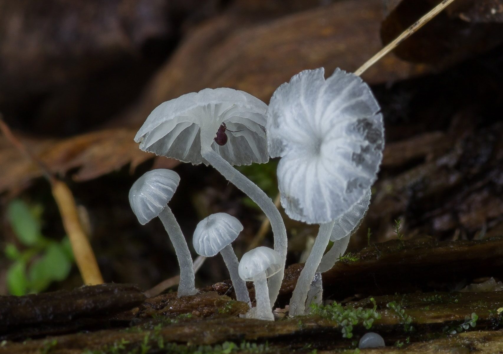
- Delicatula whole
- Delicatula young
- Whole agaricus
- Omphalia caricicola
- Mycena integrella
- Omphalia complete
- Delicatula bagnolensis
The current name is Delicatula integrella (Pers. : Fr.) Fayod 1889
The etymology of the specific epithet from delicatula, ae f, favorite. From delicatus, a, pet, itza + ulus (diminutive) and integrellus, a, um, whole, immaculate, healthy, immaculate, young. From integer, gra, grum, with the same meanings + ellus, a, um (diminutive).
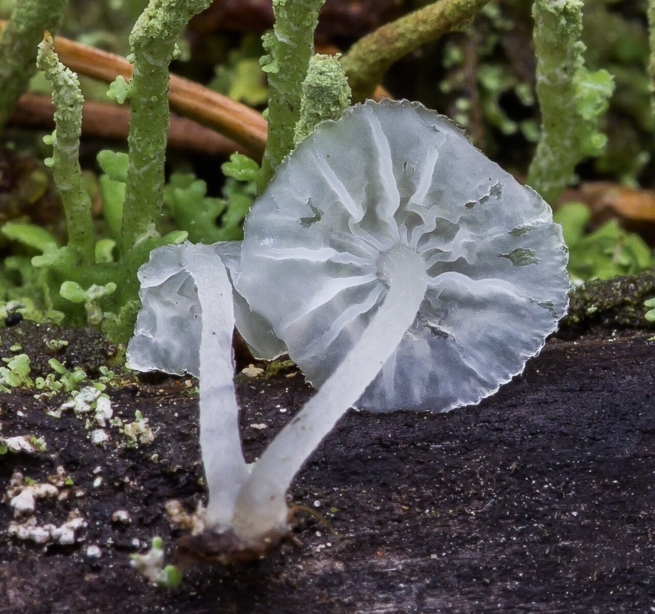
head small in size 0,3 – 1,5 cm, in young mushrooms it is hemispherical, bell-shaped, with age it becomes prostrate, “omphalino-like” with a hole in the center and opening ribbed edges. The edge itself is scalloped (serrated), uneven, in overripe specimens it may be bent upwards, and the central depression may be weakly expressed or completely absent. The surface of the cap looks smooth, hydrophobic, with radial wrinkles and translucent plates. With a slight increase (using a magnifying glass), very small villi can be seen on the surface. The color of the cap is very characteristic – pale white translucent like jelly, with age it can acquire a straw-yellow hue, especially in the center.
Hymenophore mushroom – lamellar. The plates, adnate with a tooth or slightly descending, very rare, sometimes forked, similar to veins and folds, do not reach the edge of the cap. The color is like a hat – whitish, may turn slightly yellow with age.
Pulp caps are very thin whitish, despite the gelatinous jelly-like appearance is quite durable. The flesh of the leg is more watery.
Smell and taste not expressed.
spore powder white or colorless.
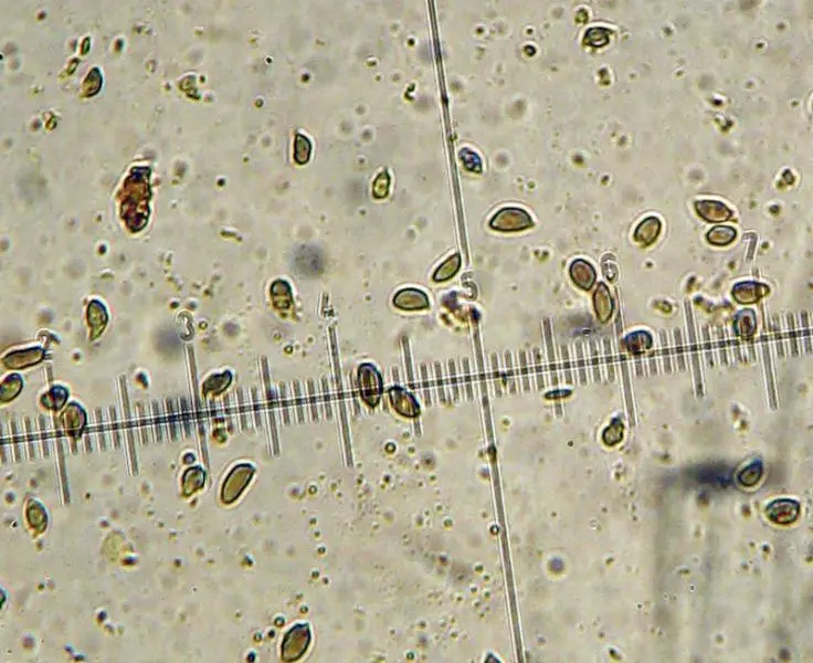
Microscopy
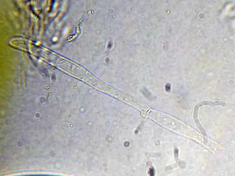
Spores 6,5–8,5 × 3,5–4,5 µm, almond-shaped to slightly fusiform, amyloid.
Observation in Meltzer’s reagent at 400× magnification:
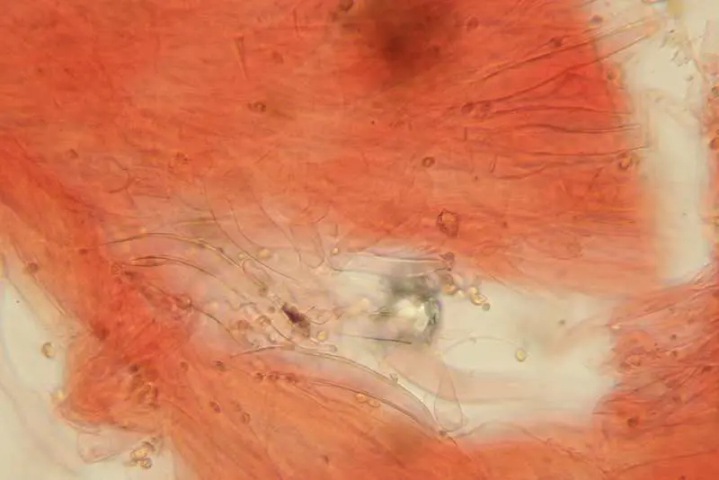
Basidia 23 – 32 (35) × 7.0 – 9.0 µm, club-shaped, 4-spored.
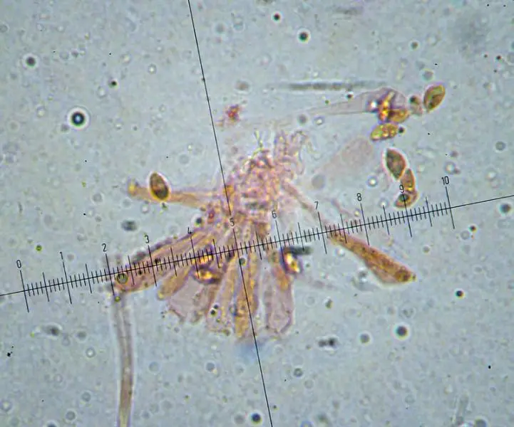
Hymenial cystidia and calocystidia are absent.
Stipitipellis is a cutis of parallel, cylindrical hyphae up to 8 (10) µm in diameter.
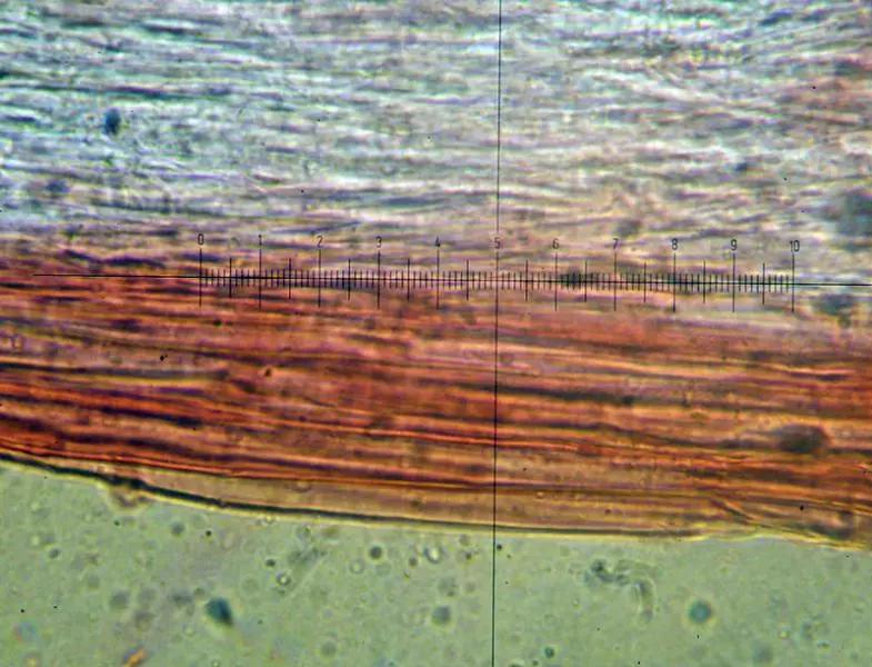
Pileipellis – cutis of radially arranged subcylindrical, thin-walled hyphae up to 10 microns in diameter.
Buckles observed:
Leg capillary-shaped, of the same color as the cap, up to 2 cm in height and up to 1,5 mm in diameter, cylindrical, often slightly curved at the base, where there is a swelling (pseudobulb). The surface is densely hairy, especially at the bottom, making the stipe appear slightly darker than the mushroom as a whole. As it matures, the stem becomes smoother and glossier.
Grows in damp areas on rotting wood, both deciduous and (rarely) coniferous trees, as well as on rotten stumps, roots, fallen branches.
May-November, with sufficient humidity after rains, it bears fruit abundantly, grows both singly and in groups. Distributed in Western Europe, the European part of Our Country, the Caucasus, Siberia, the Far East. Found in Central Asia, Africa, Australia.
The mushroom does not appear to contain poisonous substances, but is considered inedible.
It is most similar to some small mycenae with an “omphaloid” structure, but the translucent appearance and general structure of the fruiting body will make it easy to identify Delicatula small in this interesting mushroom.
Photo: Alexander Kozlovskikh, microscopy funghiitaliani.it.