


Contents
Valve tricuspide
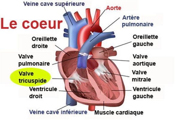
The tricuspid valve (from the Latin cusp meaning spear point, or three-pointed valve) is a valve located at the level of the heart, separating the right atrium from the right ventricle.
Tricuspid aortic valve
Position. The tricuspid valve is located at the level of the heart. The latter is divided into two parts, left and right, each having a ventricle and an atrium. The tricuspid valve separates the right atrium from the right ventricle (1).
Structure. The tricuspid valve can be divided into two parts (2):
- The valve apparatus, made up of a fibrous ring surrounding the valve and valve leaflets, originating at the level of the fibrous ring and made up of folds of the endocardium (inner layer of the heart) (1).
- The subvalvular system, made up of tendon cords and pillars called papillary muscles
Function of the tricuspid valve
Blood path. Blood circulates in one direction through the heart and the blood system. The right atrium receives venous blood, that is to say poor in oxygen and coming from the upper and lower vena cava. This blood then passes through the tricuspid valve to reach the right ventricle. Within the latter, the blood then passes through the pulmonary valve to reach the pulmonary trunk. The latter will divide into right and left pulmonary arteries to join the lungs (1).
Opening / closing of the valve. The tricuspid valve opens by pressure of the blood at the level of the right atrium. The latter contracts and allows blood to pass through the tricuspid valve to the right ventricle (1). When the right ventricle is full and the pressure increases, the ventricle contracts and causes the tricuspid valve to close. This is in particular kept closed thanks to the papillary muscles.
Anti-reflux of blood. Playing an important role in the passage of blood, the tricuspid valve also prevents backflow of blood from the right ventricle to the right atrium (1).
Valve disease: stenosis and tricuspid insufficiency
Valvular heart disease refers to all pathologies affecting the heart valves. The evolution of these pathologies can lead to a change in the structure of the heart with dilation of the atrium or the ventricle. The symptoms of these pathologies can in particular be a murmur in the heart, palpitations, or even discomfort (3).
- Tricuspid insufficiency. This pathology is linked to a poor closing of the valve leading to a back flow of blood towards the atrium. The causes of this condition are varied and may in particular be linked to acute rheumatoid arthritis, an acquired or congenital malformation, or even an infection. The latter case corresponds to endocarditis.
- Tricuspid narrowing. Rare, this valve disease corresponds to an insufficient opening of the valve preventing the blood from circulating well. The causes are varied and may in particular be related to rheumatic fever, infection or endocarditis.
Treatment of heart valve disease
Medical treatment. Depending on the valve disease and its progression, certain drugs may be prescribed for example to prevent certain infections such as infective endocarditis. These treatments can also be specific and intended for associated diseases (4) (5).
Surgical treatment. In the most advanced cases of valve disease, surgery is frequently performed. The operation consists of either repairing the valve or replacing the valve with the installation of a mechanical or biological valve prosthesis (bio-prosthesis) (3).
Examination of the tricuspid valve
Physical examination. First, a clinical examination is carried out in order to study the heart rate in particular and to assess the symptoms perceived by the patient such as shortness of breath or palpitations.
Medical imaging exam. In order to establish or confirm a diagnosis, a cardiac ultrasound, or even a doppler ultrasound can be performed. They can be supplemented by a coronary angiography, a CT scan, or an MRI.
Electrocardiogramme d’effort. This test is used to analyze the electrical activity of the heart during physical exertion.
History
Artificial heart valve. Charles A. Hufnagel, American surgeon of the 20th century, was the first to invent the artificial heart valve. In 1952, he implanted, in a patient suffering from aortic insufficiency, an artificial valve formed of a metal cage with a silicone ball placed in its center (6).