


Contents
Blood vessel
Blood vessels (vessel: from the lower Latin vascellum, from classical Latin vasculum, meaning small vessel, blood: from Latin sanguineus) are organs of blood circulation.
Anatomy
General description. Blood vessels form a closed circuit through which blood circulates. This circuit is divided into a large body circulation and a small pulmonary circulation. These vessels consist of a wall with three tunics: (1) (2)
- The inner coat, or intima, composed of a cellular layer of endothelium and lining the inner surface of the vessels;
- The middle tunic, or media, constituting the intermediate layer and composed of muscular and elastic fibers;
- The outer layer, or adventitia, constituting the outer layer and composed of collagen fibers and fibrous tissues.
Blood vessels are divided into different groups (1)
- Arteries. The arteries constitute the vessels where the blood, rich in oxygen, leaves the heart to reach the various structures of the body, except for the pulmonary and placental circulation. There are different types of arteries depending on their structure1.
– The elastic-type arteries, with a large caliber, have a thick wall and are made up of numerous elastic fibers. They are mainly localized near the heart, such as the aorta, or the pulmonary artery.
– Muscular type arteries have a smaller caliber and their wall contains many smooth muscle fibers.
– The arterioles are located at the end of the arterial network, between the arteries and the capillaries. They are usually localized in an organ and do not contain an outer coat.
- Veins. The veins are the vessels where the blood, poor in oxygen, leaves the periphery to reach the heart, except for pulmonary and placental circulation. From the capillaries, the venules, small veins, recover the blood poor in oxygen and join the veins. (1) The latter have a thinner wall than the arteries. Their wall has less elastic and muscle fibers but has a thicker outer tunic. The veins have the particularity of being able to contain more blood than the arteries. In order to facilitate venous return, the veins of the lower limbs have valves. (2)
- Veins. The veins are the vessels where the blood, poor in oxygen, leaves the periphery to reach the heart, except for pulmonary and placental circulation. From the capillaries, the venules, small veins, recover the blood poor in oxygen and join the veins. (1) The latter have a thinner wall than the arteries. Their wall has less elastic and muscle fibers but has a thicker outer tunic. The veins have the particularity of being able to contain more blood than the arteries. In order to facilitate venous return, the veins of the lower limbs have valves. (2)
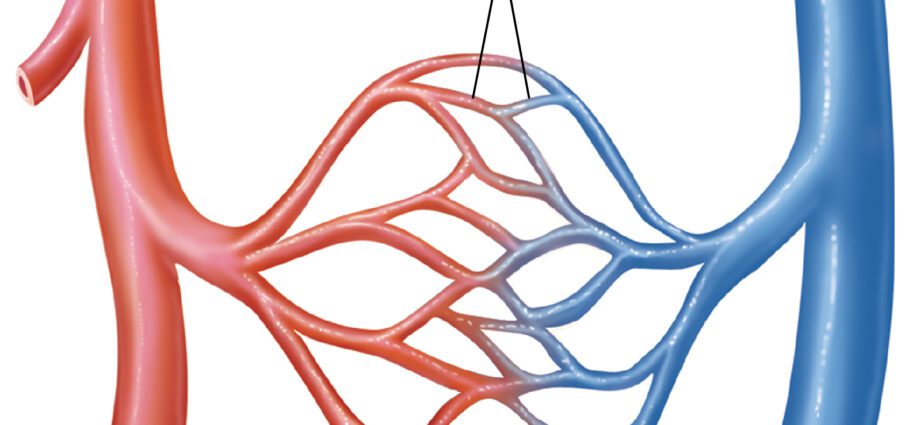
- Capillaries. Forming a branched network, capillaries are very fine vessels, with a diameter ranging from 5 to 15 micrometers. They make the transition between arterioles and venules. They allow both the distribution of oxygenated blood and nutrients; and both the recovery of carbon dioxide and metabolic waste. (1)
Innervation. The blood vessels are innervated by sympathetic nerve fibers to regulate their diameter. (1)
Functions of blood vessels
Distribution/Elimination. The blood vessels allow both the distribution of nutrients and the recovery of metabolic wastes.
Blood circulation. The blood vessels form a closed circuit. The nutrient-rich blood leaves the left ventricle of the heart through the aorta. It successively traverses the arteries, arterioles, capillaries, venules and veins. In the capillaries, the exchange of nutrients and waste takes place. The nutrient-poor blood then reaches the right atrium of the heart through the two vena cavae before enriching itself in nutrients and resuming its journey through the body. (1) (2)
Problems related to blood pressure. Too much blood pressure against the walls of the arteries can lead to high blood pressure and may increase the risk of vascular disease.3 Conversely, too low pressure leads to low blood pressure.
Thrombosis. This pathology corresponds to the formation of a blood clot in a blood vessel (4).
Stroke. Cerebrovascular accident, or stroke, is manifested by the blockage of a blood vessel in the brain, such as the formation of blood clots or the rupture of a vessel. (4)
Phlebitis. Also called venous thrombosis, this pathology corresponds to the formation of a blood clot, or thrombus, in the veins. These clots can move and move up to the inferior vena cava. This pathology can lead to various conditions such as venous insufficiency, that is to say a dysfunction of the venous network (5).
Cardiovascular illnesses. They include many pathologies such as myocardial infarction or angina pectoris. When these diseases occur, the blood vessels are often affected and can in particular cause an insufficient supply of oxygen. (6) (7)
Treatments
Drug treatments. Depending on the pathology diagnosed, certain drugs may be prescribed such as anticoagulants, anti-aggregants, or even anti-ischemic agents.
Thrombolyse. Used during strokes, this treatment consists of breaking up the thrombi, or blood clots, with the help of drugs. (5)
Surgical treatment. Depending on the pathology diagnosed and its evolution, surgery may be necessary.
Blood test
Physical examination. First, a clinical examination is carried out in order to identify and assess the pain perceived by the patient.
Medical imaging exams. X-ray, CT, MRI, coronary angiography, CT angiography, or arteriography exams can be used to confirm or deepen the diagnosis.
- Doppler ultrasound. This specific ultrasound makes it possible to observe the blood flow.
History
William Harvey, English physician of the 16th and 17th century, is known for his work and discoveries on the functioning of the blood circulation.