


Contents
Thoracic aorta
The thoracic aorta (from the Greek aortê, meaning large artery) corresponds to part of the aorta.
Anatomy
Position. The aorta is the main artery leading from the heart. It is made up of two parts:
- a thoracic part, starting from the heart and extending into the thorax, constituting the thoracic aorta;
- an abdominal part, following the first part and extending into the abdomen, constituting the abdominal aorta.
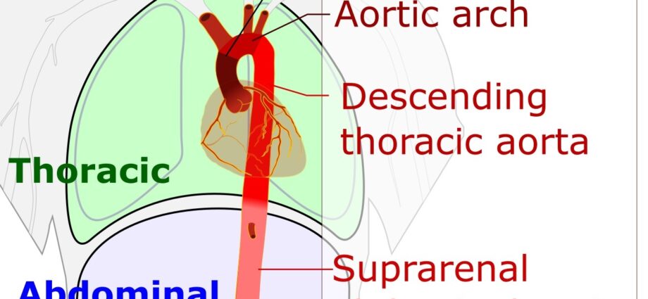
Structure. The thoracic aorta is divided into three parts (1):
- Ascending thoracic aorta. It constitutes the first part of the thoracic aorta.
Origin. The ascending thoracic aorta starts at the left ventricle of the heart.
Suitt. It goes up and has a slightly swollen appearance, called bulb of the aorta.
Termination. It ends at the level of the 2nd rib to be extended by the horizontal part of the thoracic aorta.
Peripheral branches. The ascending thoracic aorta gives rise to the coronary vessels, bound for the heart. (2)
- Horizontal thoracic aorta. Also called aortic arch or aortic arch, it is the area connecting the ascending and descending parts of the thoracic aorta. (2)
Origin. The arch of the aorta follows the ascending part, at the level of the 2nd rib.
Path. It curves and extends horizontally and obliquely, to the left and to the rear.
Termination. It ends at the level of the 4th thoracic vertebra.
Peripheral branches.
The aortic arch gives rise to several branches (2) (3):
Brachiocephalic arterial trunk. It starts at the beginning of the aortic arch, extends upwards and slightly backwards. It is divided into two branches: the right primary carotid and the right subclavian, destined for the right sternoclavicular joint.
Left primary carotid. It starts behind the aortic arch and to the left of the brachiocephalic arterial trunk. It goes up towards the base of the neck. Left subclavian artery. It starts behind the left primary carotid artery and goes up to join the base of the neck.
Neubauer’s lower thyroid artery. Inconsistent, it usually starts between the brachio-cephalic arterial trunk and the left primitive carotid artery. It goes up and ends at the thyroid isthmus.
- Descending thoracic aorta. It constitutes the last part of the thoracic aorta.
Origin. The descending thoracic aorta starts at the level of the 4th thoracic vertebra.
Path. It descends within the mediastinum, an anatomical area located between the two lungs and comprising various organs including the heart. It then passes through the diaphragmatic orifice. It continues its journey, approaching the midline to position itself in front of the spine. (1) (2)
Termination. The descending thoracic aorta terminates at the level of the 12th thoracic vertebra, and is extended by the abdominal aorta. (1) (2)
Peripheral branchess. They give rise to several branches: the visceral branches destined for the thoracic organs; the parietal branches to the chest wall.
Bronchial arteries. They start from the upper part of the thoracic aorta and join the bronchi, and their number varies.
Esophageal arteries. From 2 to 4, these fine arteries arise all along the thoracic aorta to join the esophagus.
Mediastinal arteries. Constituting small arterioles, they start on the front face of the thoracic aorta before joining the pleura, the pericardium and the ganglia.
Posterior intercostal arteries. Twelve in number, they originate on the rear face of the thoracic aorta and are distributed at the level of the corresponding intercostal spaces. (12)
Function of the thoracic aorta
Vascularization. With the help of its numerous branches supplying the thoracic wall and the visceral organs, the thoracic aorta plays a major role in the vascularization of the organism.
Wall elasticity. The aorta has an elastic wall that allows it to adapt to the pressure differences that arise during periods of cardiac contraction and rest.
Thoracic aortic aneurysm
The thoracic aortic aneurysm is congenital or acquired. This pathology corresponds to a dilation of the thoracic aorta, occurring when the walls of the aorta are no longer parallel. As it progresses, an abdominal aortic aneurysm can lead to: (4) (5)
- compression of neighboring organs;
- thrombosis, that is, the formation of a clot, in the aneurysm;
- the development of an aortic dissection;
- a fissure crisis corresponding to a “pre-rupture” and resulting in pain;
- a ruptured aneurysm corresponding to the rupture of the wall of the aorta.
Treatments
Surgical treatment. Depending on the stage of the aneurysm and the patient’s condition, surgery may be performed on the thoracic aorta.
Medical supervision. In case of minor aneurysms, the patient is placed under medical supervision but does not necessarily require surgery.
Thoracic aortic examinations
Physical examination. First, a clinical examination is performed to assess the abdominal and / or lumbar pain felt.
Medical imaging exam. In order to establish or confirm a diagnosis, an abdominal ultrasound may be performed. It can be supplemented by a CT scan, MRI, angiography, or even an aortography.
History
Neubauer’s lower thyroid artery owes its name to 18th century German anatomist and surgeon Johann Neubauer. (6)