


Symptoms of skin cancer
The first manifestations of the disease often go unnoticed. The majority of skin cancer do not cause pain, itching or bleeding.
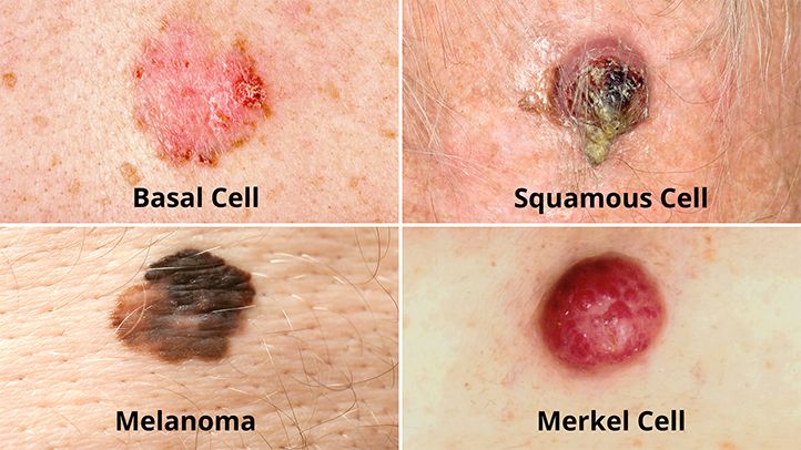
Basal cell carcinoma
70 to 80% of basal cell carcinomas are found on the face and neck and around 30% on the nose, which is the most frequent location; the other frequent locations are the cheeks, the forehead, the periphery of the eyes, in particular at the internal angle.
It is manifested in particular by one or other of the following signs:
- a flesh-colored or pinkish, waxy or “pearly” bump on the face, ears, or neck;
- a pink, smooth patch on the chest or back;
- an ulcer that does not heal.
There are four major clinical forms of basal cell carcinoma:
– Flat basal cell carcinoma or with pearly border
It is the most frequent form, forming a rounded or oval plaque, increasing in size very gradually over months or years, characterized by a pearly border (carcinomatous pearls are small growths of one to a few millimeters in diameter, firm , translucent, embedded in the skin, somewhat resembling cultured pearls, with small vessels.
– Nodular basal cell carcinoma
This frequent form also forms a translucent rise of firm consistency, waxy or pinkish white with small vessels, resembling the pearls described above. When they evolve and exceed 3-4 mm in diameter, it is common to see a depression in the center, giving them the appearance of an extinct volcano with a translucent and hilly border. They are often fragile and bleed easily.
– Superficial basal cell carcinoma
It is the only basal cell carcinoma common on the trunk (about half of cases) and limbs. It forms a pink or red plaque of slow and gradual extension.
– Basal cell carcinoma scleroderma
This basal cell carcinoma is quite rare because it represents only 2% of cases, forms a yellowish-white, waxy, hard plaque, the boundaries of which are difficult to define. Its recurrence is frequent because it is not uncommon for the ablation to be insufficient given the limits that are difficult to define: the dermatologist or the surgeon removes what he sees and there is often some left on the periphery of the operated area.
Almost all forms of basal cell carcinoma can take on a pigmented (brown-black) appearance and ulcerate when they are developed. They are then easily hemorrhagic and can initiate mutilations by destruction of the skin and the subcutaneous tissues (cartilage, bones…).
Squamous cell carcinoma
It is manifested in particular by one or other of the following signs:
- a pinkish or whitish, rough or dry patch of skin;
- a pink or whitish, firm, warty nodule;
- an ulcer that does not heal.
Squamous cell carcinoma most often develops on actinic keratosis, a small lesion rough to the touch, a few millimeters in diameter, pinkish or brown. Actinic keratoses are particularly frequent on areas exposed to the sun (convexities of the face, scalp of men with baldness, backs of the hands, forearms, etc.). People with many actinic keratoses have an approximately 10% risk of developing invasive cutaneous squamous cell carcinoma during their lifetime. The signs which should lead one to suspect the transformation of an actinic keratosis into squamous cell carcinoma are the rapid spreading of the keratosis and its infiltration (the plaque becomes more swollen and infiltrates the skin, losing its supple character to become harder) . Then, it can erode or even ulcer and sprout. This then results in a true ulcerative squamous cell carcinoma, forming a hard tumor with an irregular surface, budding and ulcerated.
Let us cite two particular clinical forms of squamous cell carcinoma:
– Bowen’s intraepidermal carcinoma: this is a form of squamous cell carcinoma limited to the epidermis, the superficial layer of the skin and therefore with little risk of metastases (the vessels allowing cancer cells to migrate are in the dermis, below the epidermis. It is most often in the form of a red, scaly patch of fairly slow development, and it is common on the legs. Lack of diagnosis leads to the risk of development in infiltrating squamous cell carcinoma.
– Keratoacanthoma: it is a rapidly appearing tumor, frequent on the face and the top of the trunk, resulting in a “stuffed tomato” apsect: central horny zone with pinkish white rim with vessels.
Melanoma
Un normal mole is brown, beige or pinkish. It is flat or raised. It is round or oval, and its outline is regular. It measures, most of the time, less than 6 mm in diameter, and above all, it does not change. |
It is manifested in particular by one or other of the following signs.
- a mole that changes color or size, or has an irregular outline;
- a mole that is bleeding or has areas of red, white, blue, or blue-blackish color;
- a blackish lesion on the skin or on a mucous membrane (for example, the mucous membranes of the nose or mouth).
Remark. Melanoma can occur anywhere on the body. However, it is found most often on the back in men, and on one leg in women.