


Contents
What is retinal detachment
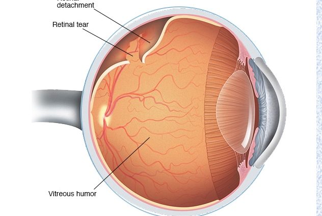
– Retinal detachment is a disease that leads to decreased vision and even loss of vision. It can occur either due to a rupture of the retina, under which the intraocular fluid begins to flow, or as a result of a traction syndrome, when there is a growth between the vitreous body and the retina, and the vitreous body begins to pull, resulting in such a detachment. Also, retinal detachment can occur if there is a hemorrhage under it, a tumor is already a secondary detachment, says Candidate of Medical Sciences, ophthalmologist of the highest category Natalya Voroshilova.
As the doctor explained, detachment can be primary and secondary. Primary pathology is called, in which detachment is preceded by a rupture, followed by leakage of fluid under the retina and detachment of this most important membrane of the eye. Secondary detachment develops as a complication of any pathological process – for example, due to the appearance of a neoplasm between the retina and vascular membranes of the eye.
There are several types of fiber detachment:
- rhematogenous (means rupture) – it occurs due to a rupture of the retina;
- traction – occurs due to the tension of the retinal tissue from the side of the vitreous body;
- exudative – occurs when serous fluid penetrates into the space under the retina, and vascular permeability increases;
- mixed – for example, traction-rhegmatogenous type, in which the gap is formed against the background of traction of the vitreous body.
Causes of retinal detachment
The main cause of the disease is the rupture of the retina. Through the gap formed, fluid from the vitreous body penetrates under the retina and exfoliates it from the choroid. That is, there is a traction of the vitreous body when its normal state changes.
Retinal breaks can also occur when it is thinned. Large tears often occur with eye injuries. Ophthalmologists also note that fiber detachment can occur even in people with excellent vision and in those who have never had eye problems. The reasons may be excessive physical exertion and strong shaking of the body during jumps and falls. It is recommended for people with excellent physical data and vision not to miss preventive appointments with an ophthalmologist and to be attentive to the health of their eyes.
Symptoms of retinal detachment
At first, the disease in a person is asymptomatic, in the future, retinal detachment of the eye can be indicated by:
- the appearance of a “veil” before the eye;
- flashes in the form of sparks and lightning;
- distortion of the considered letters, objects, falling out of the field of view of their individual sections.
Some patients also note that vision deteriorated after sleep. The fact is that with a horizontal position of the body, the retina returns to its place, and when a person stands up, that is, takes a vertical position, it again moves away from the choroid and visual defects resume.
Treatment of retinal detachment
Unfortunately, no magic pills and drops can cure retinal detachment. The only option left is surgery. According to doctors, the sooner the operation is performed, the more likely it is to restore vision and save the eye.
During the operation, the surgeon will have to detect a retinal tear, close it and create a strong adhesion between the vascular and retinal membranes.
Diagnostics
To diagnose retinal detachment, you should definitely consult an ophthalmologist. The doctor will check the visual acuity, examine the field of view, conduct a special electrophysiological study to determine the viability of the nerve cells of the retina and optic nerve. If necessary, you can also conduct a study using ultrasound to determine the size of the detached retina and the condition of the vitreous body and examine the fundus (ophthalmoscopy) to accurately determine the location of retinal breaks and their number.
Only after the results have been carried out, the doctor will be able to say which surgical intervention is suitable for the patient.
Modern treatments
There are several types of surgery, the doctor will choose one of them depending on the specific type of detachment.
- Local filling. It is carried out in the zone of retinal rupture in those cases when it has detached partially;
- Circular filling. It is used in more severe cases when the retina has detached completely and there are multiple breaks;
- Vitrectomy. This is a method in which the altered vitreous body is removed from the eye and one of the necessary drugs is injected instead: saline, liquid silicone, a perfluorocarbon compound in the form of a liquid, or a special gas that presses the retina against the choroid from the inside;
- Laser coagulation or cryopexy to limit the area of rupture and thinned areas of the retina;
- Retinopexy. It is performed using special sapphire micronails to fix the torn edge of the retina in case of its giant breaks.
Prevention of retinal detachment at home
Retinal detachment is a dangerous complication of myopia, as well as age-related or hereditary circulatory disorders of the eye. The only way to prevent the disease is to consult a doctor in time for complaints and not to miss preventive examinations.
It is also worth noting that even after surgical treatment of retinal detachment, relapses may occur. If you have already encountered such a problem and do not want to meet again, then you need to conduct a thorough examination of the retina through a wide pupil by a specialist using special equipment and, if necessary, preventive laser coagulation of the retina.
Ophthalmologists also advise pregnant women to be observed by doctors – for the entire pregnancy at least twice, at the beginning and end of pregnancy. After the birth of a child, the mother should be examined by an ophthalmologist no later than 1-3 months after them.
Popular questions and answers
Comments Natalia Voroshilova, PhD, ophthalmologist of the highest category: