


Contents
Ovarian follicle
Ovarian follicles are structures located within the ovaries and involved in ovulation.
Anatomy of the ovarian follicle
Position. Ovarian follicles are located in the cortical area of the ovaries. Two in number, the female ovaries or gonads are glands located in the small pelvis, at the back of the uterus1. They also adjoin the fallopian tubes, whose fringes border them to form a pavilion. Ovoid in shape and 3 to 4 cm long, the ovaries consist of 2 parts:
- At the periphery of the ovary is the cortical zone, where the ovarian follicles are located;
- In the center of the ovary is the spinal cord area, which is made up of connective tissue and blood vessels.
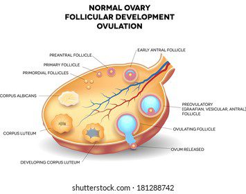
Structure. Each ovarian follicle contains an oocyte, which will then become the ovum. The structure of ovarian follicles varies according to their stage of maturation (2) (3):
- Primordial follicle: It designates an ovarian follicle whose maturation has not yet started. This type of follicle corresponds to the one found mainly in the cortical area.
- Primary follicle: It corresponds to the first stage of maturation of the follicle where the oocyte and the cells surrounding it grow.
- Secondary follicle: At this stage, several layers of epithelium form around the oocyte. The latter also continues to grow. The follicular cells then take the name of granular cells.
- Mature secondary follicle: A layer of cells develops around the follicle, forming the follicular theca. At this stage, the oocyte secretes a substance forming a thick membrane, the zona pellucida. A translucent liquid also collects between the granular cells.
- Mature ovarian follicle or De Graaf’s follicle: The fluid accumulated between the granular cells groups together and forms a cavity, the follicular antrum. As it continues to fill with fluid, the cavity grows to finally isolate the oocyte surrounded by its cell capsule, called corona radiata. When the follicle reaches its maximum dimensions, it is ready for ovulation.
- Corpus luteum: During ovulation, the oocyte is expelled while the follicle collapses. The granular cells multiply to fill the space left by the oocyte. These cells transform and become luteal cells, giving rise to a follicle called the corpus luteum. The latter has an endocrine function by synthesizing in particular progesterone, a hormone involved in the event of fertilization of the ovum.
- White body: This last stage corresponds to the total degeneration of the follicle.
Ovarian cycle
Lasting an average of 28 days, the ovarian cycle refers to all the phenomena allowing the maturation of an egg within the ovary. These phenomena are controlled by different hormonal processes and are divided into two phases (2) (3):
- Follicular phase. It takes place from the 1st to the 14th day of the ovarian cycle and ends during ovulation. During this phase, several primordial ovarian follicles begin to mature. Only one of these ovarian follicles reaches the De Graaf follicle stage and corresponds to the follicle responsible for the expulsion of the oocyte during ovulation.
- Luteal phase. It takes place from the 14th to the 28th day of the cycle and corresponds to the degeneration of the follicle. During this period, the ovarian follicles evolve into yellow bodies then white.
Pathology and disease of the ovary
Ovarian cancer. Malignant (cancerous) or benign (non-cancerous) tumors can appear in the ovary, where the ovarian follicles are located (4). Symptoms can include pelvic discomfort, menstrual cycle problems or pain.
Ovarian Cyst. It corresponds to the development of a pocket outside or inside the ovary. The structure of the ovarian cyst is variable. Two categories of cysts are distinguished:
- The most common functional cysts resolve spontaneously (1).
- Organic cysts need to be taken care of as they can lead to discomfort, pain, and in some cases the development of cancer cells.
Treatments
Surgical treatment. Depending on the pathology diagnosed and its evolution, a surgical intervention can be implemented as a laparoscopic surgery in certain cases of ovarian cysts.
Chemotherapy. Depending on the type and stage of cancer, treatment of the tumor may be accompanied by chemotherapy.
Examination of the ovaries
Physical examination. First, a clinical examination is performed to identify and assess the symptoms perceived by the patient.
Medical imaging exam. Depending on the suspected or proven pathology, additional examinations may be performed such as an ultrasound or an x-ray.
Laparoscopy. This examination is an endoscopic technique allowing access to the abdominal cavity, without opening the abdominal wall.
Biological examination. Blood tests can be performed to identify, for example, certain tumor markers.
History
Originally, the ovaries designated only the organs where eggs are formed in oviparous animals, hence the Latin etymological origin: ovum, egg. The term ovary was then assigned by analogy to the female gonads in viviparous animals, which were then referred to as the female testes (5).