


Mollet
The calf (from Old French soft, soft) is a fleshy area located on the posterior part of the leg, between the back of the knee and the ankle.
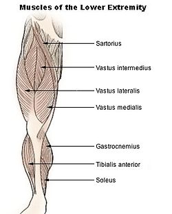
Calf Anatomy
Shape and structure. The calf owes its shape and structure to the muscles that compose it and which come from the posterior and external compartment of the leg.
Musculature. Located in the posterior compartment, the triceps sural muscle gives shape to the calf. The sural triceps muscle is made up of three bundles: the lateral gastrocnemius, the medial gastrocnemius and the solar muscle, located under the two gastrocnemius. (1) The sural triceps terminates in the Achilles tendon. Two muscles of the external compartment also make up the calf: the lateral fibular long and the lateral fibular short.
Vascularization and innervation. The triceps sural muscle is innervated by the tibial nerve (2). The muscles of the external compartment are innervated by the superficial peroneal nerve. (3) The whole is vascularized by the posterior tibial and fibular arteries.
Calf functions
Plantar flexion. The calf muscles are involved in plantar flexion of the ankle. (2)
Eversion of the foot. The muscles of the external compartment are responsible for the eversion of the foot, that is to say for the movement bringing the plantar face outwards.
Stabilization of the foot. The role of the muscles of the external compartment is to stabilize the foot, especially during plantar flexion. (4)
Calf pathologies
Tendinopathies. They designate all the pathologies that can occur in the tendons. They are mainly manifested by pain during exertion. The causes of these pathologies can be varied. The origin can be intrinsic as well with genetic predispositions, as extrinsic, with for example bad positions during the practice of sport (5).
Muscle pain without lesions
- Contracture. It is an involuntary, painful and permanent contraction of a muscle.
- Cramp. It corresponds to an involuntary, painful and temporary contraction of a muscle.
Muscle injury. The calf can be the site of muscle damage, accompanied by pain.
- Elongation. First stage of muscle damage, elongation corresponds to a stretching of the muscle caused by microtears and resulting in muscle disorganization.
- Breakdown. Second stage of muscle damage, the breakdown corresponds to a rupture of muscle fibers.
SOON BACK. The last stage of muscle damage, it corresponds to a total rupture of a muscle.
Varicose veins. This pathology corresponds to the abnormal dilation of the veins. Affecting the superficial venous network of the lower limbs, varicose veins are visible on the surface of the calf. They are often associated with pain and heaviness felt in the legs.
Calf prevention and treatment
Drug treatments. Depending on the pathology diagnosed, different treatments may be prescribed to reduce pain and inflammation.
Symptomatic treatment. In the case of varicose veins, elastic compression may be prescribed to decrease the dilation of the veins.
Endovascular treatment. This is a treatment performed within the blood vessels.
Surgical treatment. Depending on the type of pathology diagnosed, surgery may be performed.
Physical treatment. Physical therapies, through specific exercise programs, can be prescribed such as physiotherapy or physiotherapy.
Calf exams
Physical examination. First, a clinical examination is performed in order to observe and assess the symptoms perceived by the patient.
Medical imaging examination. X-ray, CT, or MRI exams can be used to confirm or further the diagnosis.
Doppler ultrasound. This specific ultrasound makes it possible to observe the blood flow. It is used in particular to diagnose varicose veins.