


Contents
Lobe occipital
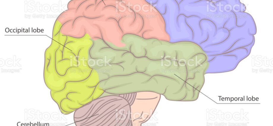
The occipital lobe (lobe – from the Greek lobos, occipital – from the medieval Latin occipitalis, from occiput) is one of the regions of the brain, located laterally and at the back of the brain.
Anatomy
Position. The occipital lobe is located at the level of the occipital bone, on the lateral and lower part of the brain. It is separated from the other lobes by different grooves:
- The occipito-temporal sulcus separates it from the temporal lobe located in front.
- The parieto-occipital groove separates it from the parietal lobe located above and in front.
- The calcarin groove is located below the occipital lobe.
Main structure. The occipital lobe is one of the regions of the brain. The latter is the most developed part of the brain and occupies most of it. It is made up of neurons, the cell bodies of which are located on the periphery and form the gray matter. This outer surface is called the cortex. The extensions of these bodies, called nerve fibers, are located in the center and form the white matter. This internal surface is called the medullary region (1) (2). Numerous furrows, or cracks when they are deeper, distinguish different areas within the brain. The longitudinal fissure of the brain allows it to be separated into two hemispheres, left and right. These hemispheres are connected to each other by commissures, the main one of which is the corpus callosum. Each hemisphere is then divided, through the primary sulcus, into four lobes: the frontal lobe, the parietal lobe, the temporal lobe and the occipital lobe (2) (3).
Structure du lobe occipital. The occipital lobe has secondary and tertiary grooves, making it possible to form convolutions called gyri.
Features
The cerebral cortex is associated with mental, sensitivomotor activities, as well as the origin and control of skeletal muscle contraction. These different functions are distributed in the different lobes of the brain (1).
Function of the occipital lobe. The occipital lobe essentially has somatosensory functions. It includes the center of vision (2) (3).
Pathologies associated with the occipital lobe
Of degenerative, vascular or tumor origin, certain pathologies can develop in the occipital lobe and affect the central nervous system.
Stroke. Cerebrovascular accident, or stroke, occurs when a blood vessel is blocked in the brain, such as blood clots or a ruptured vessel (4). This pathology can impact the functions of the occipital lobe.
Head trauma. It corresponds to a shock at the level of the skull which can cause brain damage, in particular at the level of the occipital lobe. (5)
Multiple sclerosis. This pathology is an autoimmune disease of the central nervous system. The immune system attacks the myelin, the sheath surrounding nerve fibers, causing inflammatory reactions. (6)
Tumor of the occipital lobe. Benign or malignant tumors can develop in the brain, especially in the occipital lobe. (7)
Degenerative cerebral pathologies. Certain pathologies can lead to changes in nervous tissue in the brain.
- Alzheimer’s disease. It results in a modification of cognitive faculties with in particular a loss of memory or reasoning. (8)
- Parkinson disease. It is manifested in particular by a tremor at rest, a slowing down and a reduction in the range of motion. (9)
Treatments
Drug treatments. Depending on the pathology diagnosed, certain treatments may be prescribed such as anti-inflammatory drugs.
Thrombolyse. Used during strokes, this treatment consists of breaking up the thrombi, or blood clots, with the help of drugs. (4)
Surgical treatment. Depending on the type of pathology diagnosed, surgery may be performed.
Chemotherapy, radiotherapy, targeted therapy. Depending on the stage of the tumor, these treatments can be implemented.
Brain exam
Physical examination. First, a clinical examination is performed in order to observe and assess the symptoms perceived by the patient.
Medical imaging exam. In order to establish or confirm a diagnosis, a cerebral and spinal CT scan or a cerebral MRI may in particular be performed.
biopsy. This examination consists of a sample of cells.
Lumbar puncture. This exam allows the cerebrospinal fluid to be analyzed.
History
Louis Pierre Gratiolet, French anatomist of the 19th century, is one of the first to have introduced the principle of divisions of the cortex into lobes.