


Contents
Humerus
The humerus (from Latin humerus) is the only bone in the arm located between the shoulder and the elbow.
Anatomy of the humerus
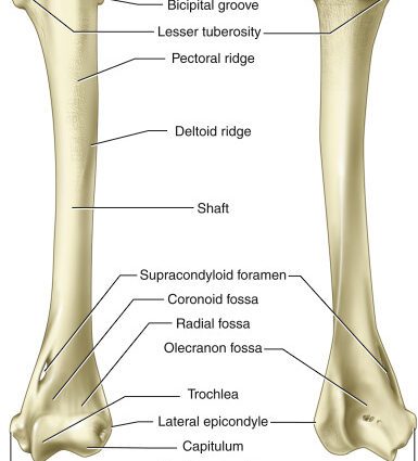
Overall structure. Among the bones of the upper limb, the humerus is the longest and most voluminous bone. (1) Elongated in shape, this bone is made up of three parts:
- a proximal end, located at the level of the shoulder, which articulates with the scapula (or scapula).
- a distal end, located at the elbow, which articulates with the radius and ulna (or ulna).
- a diaphysis, central part of the bone located between the two ends.
Joints. The proximal end of the humerus forms the head of the humerus, hemispherical in appearance, which is anchored in the cavity of the scapula (or scapula).
The distal end is composed of two condyles or articular surfaces: the trochlea and the capitulum articulating respectively with the ulna (or ulna) and the radius. Three pits also make up the distal end, where the ulna and radius are anchored during flexion and extension movements of the elbow.
Insertion of muscles and ligaments. The proximal end of the humerus is made up of two tubercles, major and minor, as well as an intertubercular groove. The set serves as attachment points for muscles and tendons.
In the center of the shaft is the deltoid tuberosity which serves as an attachment point for the deltoid muscle of the shoulder.
The distal end is made up of two epicondyles, medial and lateral, bony protrusions serving as points of attachment to muscles and ligaments.
Humerus physiology
Forearm movements. Anchored at the level of the humerus, the arm muscles are responsible for the various movements of the forearm such as flexion, extension, supination or even abduction. (2)
Arm movements. Anchored at the level of the humerus, the muscles of the shoulder joint allow movement of the arm.
Joint movements. The muscles anchored in the humerus participate in the mobility of the elbow and wrist joints.
Pathologies of the humerus
The humerus can fracture. These can occur at the level of the shaft (central part of the humerus), the lower extremity (elbow), or the upper extremity (shoulder). A fracture in the upper extremity may be accompanied by a dislocation of the shoulder (3).
Bone diseases.
- Osteoporosis. This pathology constitutes a loss of bone density which is generally found in people over the age of 60. It accentuates bone fragility and promotes bills. (4)
- Bone cancer. Metastases can develop in the bones. These cancer cells usually originate from primary cancer in another organ. (5)
- Bone dystrophy. This pathology constitutes an abnormal development or remodeling of bone tissue and includes many diseases. One of the most common, Paget’s disease (6) causes bone densification and deformation, leading to pain. As for algodystrophy, it corresponds to the appearance of pain and / or stiffness following a trauma (fracture, surgery, etc.).
Treatments and prevention of the humerus
Drug treatments. Depending on the disease, different treatments may be prescribed to regulate or strengthen bone tissue, as well as to reduce pain and inflammation.
Surgical treatment. Depending on the type of fracture, a surgical operation can be performed with the placement of pins, a screw plate, an external fixator or in some cases a prosthesis.
Orthopedic treatment. Depending on the type of fracture, the installation of a plaster or a resin can be carried out.
Physical treatment. Physical therapies, such as physiotherapy or physiotherapy, may be prescribed.
Hormonal treatment, radiotherapy or chemotherapy. These treatments may be prescribed depending on the stage of cancer progression.
Humerus examinations
Physical examination. Diagnosis begins with an assessment of arm pain to identify its causes.
Medical imaging examination. Depending on the suspected or proven pathology, additional examinations may be performed such as an X-ray, an ultrasound, a CT scan, an MRI, a scintigraphy or even a bone densitometry.
Medical analysis. In order to identify certain pathologies, blood or urine analyzes can be carried out such as, for example, the dosage of phosphorus or calcium.
Bone biopsy. In some cases, a bone sample is taken to confirm a diagnosis.
History and symbolism of the humerus
In 2014, the National Center for Scientific Research (CNRS) as well as the National Institute of Preventive Archeology unveiled the discovery of arm and forearm bones, including that of the humerus, on the banks of the Seine. in Normandy. These bones belonged to a pre-Neanderthal ancestor dating back 200 years (000).