


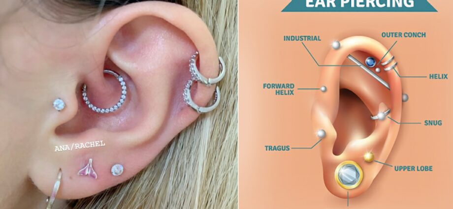
Helix
The helix (from the scientific Latin helix, from the Greek heliks, -ikos, meaning spiral) is a structure of the outer ear.
Anatomy
Position. The helix forms the upper and lateral border of the auricle, or auricular pinna. The latter corresponds to the visible part of the outer ear while the external acoustic meatus represents the invisible part. The auricle, or pinna, is thus referred to in everyday language as the ear, although the latter is actually made up of three parts: the outer ear, the middle ear and the inner ear (1).
Structure. The helix corresponds to the upper and lateral part of the outer ear. The latter is mainly composed of elastic cartilage lined with a thin layer of skin, as well as fine and sparse hairs. Unlike the helix, the lower part of the outer ear, called the lobule, is a fleshy part devoid of cartilage (1).
Vascularization. The helix and its root are supplied by the upper and middle anterior atrial arteries, respectively (2).
Helix functions
Auditory role. The auricle, or pinna, plays a role in hearing by collecting and amplifying sound frequencies. The process will continue in the external acoustic meatus and then in the other parts of the ear.
Label this text field
Pathology and associated issues
Text
Acouphènes. Tinnitus corresponds to abnormal noises perceived in a subject in the absence of external sounds. The causes of this tinnitus are varied and can in some cases be linked to certain pathologies or linked to cellular aging. Depending on the origin, duration, and associated problems, tinnitus is divided into several categories (3):
- Objective and subjective tinnitus: Objective tinnitus corresponds to a physical sound source coming from inside the subject’s body, such as for example a blood vessel. For subjective tinnitus, no physical sound source is identified. It corresponds to a bad processing of sound information by the auditory pathways.
- Acute, subacute and chronic tinnitus: They are distinguished according to their duration. Tinnitus is said to be acute when it lasts for three months, subacute for a period of between three and twelve months and chronic when it lasts for more than twelve months.
- Compensated and decompensated tinnitus: They define the impact on the quality of life. Compensated tinnitus is considered “surmountable” on a daily basis, while decompensated tinnitus becomes really harmful to daily well-being.
Hyperacousie. This pathology corresponds to a hypersensitivity of sounds and external noises. It causes daily discomfort for the patient (3).
Microtie. It corresponds to a malformation of the helix, linked to insufficient development of the pinna of the ear.
Treatments
Medical treatment. Depending on the pathology diagnosed, certain drug treatments may be prescribed.
Surgical treatment. Depending on the pathology diagnosed, a surgical operation may be performed.
Examination of the helix
Physical examination. First, a clinical examination is performed in order to identify and assess the symptoms perceived by the patient.
ENT imaging exam. Tympanoscopy or nasal endoscopy may be done to confirm a diagnosis.
Symbolic
Aesthetic symbol. In different cultures, the auricular pinna of the ear is associated with an aesthetic symbol. Artificial additions are placed on the helix in particular, such as piercings.