


Contents
In line with its mission, the Editorial Board of MedTvoiLokony makes every effort to provide reliable medical content supported by the latest scientific knowledge. The additional flag “Checked Content” indicates that the article has been reviewed by or written directly by a physician. This two-step verification: a medical journalist and a doctor allows us to provide the highest quality content in line with current medical knowledge.
Our commitment in this area has been appreciated, among others, by by the Association of Journalists for Health, which awarded the Editorial Board of MedTvoiLokony with the honorary title of the Great Educator.
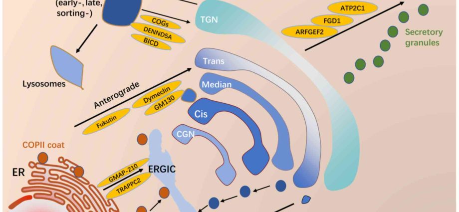
The Golgi apparatus is an organelle found in most eukaryotic cells and transports molecules from the endoplasmic reticulum to their destination. It consists of a pile of flattened tanks. There are two forms of the Golgi apparatus, the reticulate, which is characteristic of vertebrate cells (not only in oocytes and sperm), and the scaly or dictiosomal, which occurs in plant cells and in invertebrate cells.
What is the Golgi apparatus?
The Golgi apparatus is located between the endoplasmic reticulum and the cell membrane and often appears to be an extension of the endoplasmic reticulum, which is slightly smaller and smoother. The Golgi apparatus supplies different cell products to different organelles throughout the cell. The Golgi apparatus is also important in labeling products with proteins and sugar molecules that serve as identifiers so that they can be delivered to their intended destination.
Most often, cell products and proteins are produced in the endoplasmic reticulum, which has several ribosomes that make up proteins from the instructions contained in RNA. In the endoplasmic reticulum, protein products are modified, and as they reach the Golgi apparatus, more modifications are made. Eventually, the products are packaged and labeled with other proteins and molecules. Packets are released and, based on their tags or labels, are transferred to the appropriate place in the cell through the cytoskeleton.
See also: Human genetic code – its tasks and characteristics
Golgi apparatus – construction
The Golgi apparatus is made of flat pouches that fit together to form organelles, they are called by tankers. It is worth adding that in most organisms there are from four to eight such tanks, but sometimes there can be up to 60 tanks in one Golgi apparatus. The spaces between the cisterns are the light of the Golgi apparatus.
The Golgi apparatus is divided into three parts:
- cisterns near the endoplasmic reticulum, i.e. yew pages;
- cisterns on the opposite side of the endoplasmic reticulum, i.e. trans pages;
- middle tanks.
The cis side receives the packet sent from the endoplasmic reticulum by special transporters called vesicles. The opposite side, called the trance side, is responsible for transport. In addition, it should be noted that enzymes also play a huge role in the Golgi apparatus. They reside in cisterns and allow lipids and proteins to be modified as they pass from the cis surface through the medial compartment on the way to the trans compartment.
The modifications made by the various enzymes in the tank bags have a great influence on the results of the modified biomolecules. In other cases, the modifications act as labels that inform the Golgi shipping center of the final destination of the biomolecules.
There are structural and organizational differences in the Golgi apparatus among eukaryotes. In some yeasts, golgi stacking is not observed. Shepherd’s figs arranged the Golgi apparatus while Saccharomyces cerevisiae no. In plants, the individual piles of the Golgi apparatus appear to function independently.
The Golgi apparatus is usually larger and more numerous in cells that synthesize and secrete large amounts of substances; for example, antibody-secreting plasma B cells of the immune system have significant golgi apparatus.
Golgi apparatus – functions
The Golgi apparatus is the main “station” for collecting and sending protein products obtained from the endoplasmic reticulum. Proteins synthesized in the endoplasmic reticulum are packed into vesicles, which then bind to the Golgi apparatus. These proteins are modified and intended to be secreted by exocytosis or to be used in a cell. In this regard, the Golgi apparatus can be considered similar to a post office: it packs and labels items, which it then sends to different parts of the cell or to the extracellular space. The Golgi apparatus is also involved in lipid transport and the formation of lysosomes.
The structure and function of the Golgi apparatus are closely related. The individual cisterns have different enzyme assortments, allowing the proteins to be processed progressively as they move from the cisterns to the trans side. The enzymatic reactions in the cysteranes only take place near the surface of the membrane, where the enzymes are anchored. Unlike the endoplasmic reticulum, which has soluble proteins and enzymes in its lumen. A large part of enzymatic processing is post-translational modification of proteins.
For example, phosphorylation of oligosaccharides on lysosomal proteins occurs at the beginning of the cis side. The Cis side is associated with the removal of mannose residues. Removal of residual mannose and addition of N-acetylglucosamine takes place in the middle cisterns. The addition of galactose and sialic acid occurs in the trans sides. Sulphation of tyrosine and carbohydrates also takes place in them. Other general post-translational modifications to proteins include the addition of carbohydrates (glycosylation) and phosphates (phosphorylation).
Protein modifications can create a signal sequence that determines the final destination of the protein. For example, the Golgi apparatus adds a mannose-6-phosphate tag to proteins destined for lysosomes. Another important function of the Golgi apparatus is the formation of proteoglycans. The enzymes in the Golgi apparatus attach proteins to glycosaminoglycans, thereby forming proteoglycans. Glycosaminoglycans are long unbranched polysaccharide molecules present in the extracellular matrix of animals.
See also: What is Galactosemia?
Golgi apparatus in Alzheimer’s disease
Inside our cells there are many extremely complex and still relatively poorly understood structures. Cells synthesize a large number of different macromolecules. The Golgi apparatus is an integral part of modifying, sorting and transporting these macromolecules through the release of metabolites that are produced inside the cell to the outside (exocytosis) or used in the cell. The Golgi apparatus mainly modifies proteins delivered from the rough endoplasmic reticulum, but it is also involved in the transport of lipids around the cell and the formation of lysosomes.
In the brain cells of Alzheimer’s patients, the Golgi apparatus appears to break down, and researchers suggest that this is an important step in the progression of pathological effects at the biochemical level. Scientists have identified one of the mechanisms by which the progression of Alzheimer’s disease sabotages the structures of the Golgi apparatus. They blocked it and consequently observed a reduction in levels of the characteristic harmful beta-amyloid associated with Alzheimer’s disease. This is quite promising, even if it is only achieved in cell research and not in laboratory animals.
Scientists at the University of Michigan say understanding this mechanism helps decode the build-up of amyloid plaques in the brains of Alzheimer’s patients – plaques that kill cells and contribute to memory loss and other symptoms of Alzheimer’s disease.
Scientists have discovered the molecular process responsible for the fragmentation of the Golgi apparatus and have developed two techniques to ‘rescue’ the Golgi structure and hope to use them as a strategy for delaying disease progression.
It has been found that accumulation of Abeta peptide – a major factor in the formation of cell killing plaques in the brain of Alzheimer’s patients – triggers fragmentation of the Golgi apparatus by activating an enzyme called cdk5 that modifies Golgi structural proteins such as GRASP65. The scientists ‘saved’ the Golgi apparatus in two ways: they either inhibited the cdk5 enzyme or expressed the protein GRASP65, which cannot be modified by cdk5. Both rescue measures reduced harmful Abeta shedding by about 80 percent. The next step is to see if fragmentation of the Golgi can be delayed or reversed in mice.
Some studies in the field of Alzheimer’s disease suggest that brain amyloid levels are quite dynamic, and therefore Alzheimer’s disease may very well be the progressive failure of processes that work to remove harmful amyloid rather than the slow accumulation of unwanted compounds. If this is the case, a way to slow down the rate of formation may be enough to bring the sick person back to a relatively healthy state. The only solution in the long run, however, is to identify and eliminate the root causes of the disease, no matter what they may be.
See also: Caffeine will protect against Alzheimer’s?