


Contents
Electromyogramme
A benchmark examination in neurology, the electromyogram (EMG) makes it possible to analyze the electrical activity of nerves and muscles. In addition to the clinical examination, it helps in the diagnosis of various nervous and muscular pathologies.
What is the electromyogram?
The electromyogram, also called electroneuromyogram, electronography, ENMG or EMG, aims to analyze nerve impulses in motor nerves, sensory nerves and muscles. Key examination in neurology, it allows to evaluate the functioning of nerves and muscles.
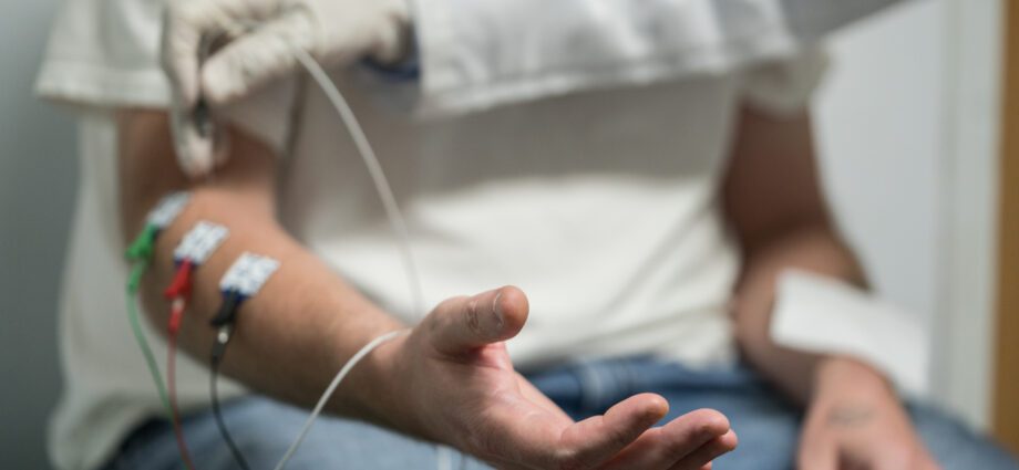
In practice, the examination consists in recording the electrical activity of the nerves as well as the contraction of a muscle either by sticking a needle in the muscle or next to the nerve, or by sticking an electrode on the skin if the nerve or the muscle are superficial. The electrical activity is analyzed at rest, after artificial electrical stimulation or by voluntary contraction effort of the patient.
How does an electromyogram work?
The examination is carried out in the hospital, in the laboratory for the functional exploration of the nervous system, or in the office of the neurologist if it is equipped. No preparation is necessary. The examination, without risk, lasts 45 to 90 minutes depending on the protocol used.
The device for performing EMG is called an electromyograph. Using electrodes (small patches) placed on the skin, it stimulates the nerve fibers electrically by sending very brief (from a tenth to a millisecond) and low intensity (a few thousandths of an ampere) electric shocks. ). This nerve current is propagated to the muscle, which will then contract and move. Sensors glued to the skin make it possible to record the electrical activity of the nerve and / or muscle. This is then transcribed on the device and analyzed on screen in the form of plots.
Depending on the symptoms and the pathology sought, different types of tests can be used:
- the actual electromyogram consists of studying the electrical activity of the muscle at rest and when the patient voluntarily contracts it. It is possible to study the activity of only a few muscle fibers. For this, the doctor introduces a fine needle, with a sensor, inside the muscle. The analysis of the electrical activity of the muscle makes it possible to detect a loss of motor nerve fibers or an abnormality of the muscle;
- the study of the conduction speeds of the motor fibers consists in stimulating the nerve at two points in order to analyze the speed and the conduction capacities of the nerve impulses on the one hand, and the muscular response on the other hand;
- the study of sensory conduction speeds makes it possible to measure the conduction of the sensory fibers of the nerve to the spinal cord;
- repetitive stimulation tests are used to test the reliability of the transmission between the nerve and the muscle. The nerve is repeatedly stimulated and the muscle response is analyzed. In particular, it is checked that its amplitude does not decrease abnormally with each stimulation.
Electrical stimulation can be more unpleasant than painful. The fine needles can cause very slight pain.
When to have an electromyogram?
The electromyogram can be prescribed in the face of different symptoms:
- after an accident that may have resulted in nerve damage;
- muscle pain (myalgia);
- muscle weakness, loss of muscle tone;
- persistent tingling, numbness, tingling (paramnesia);
- difficulty urinating or holding urine, passing or holding stool
- erectile dysfunction in men;
- unexplained perineal pain in women.
Electromyogram results
Depending on the results, the examination can diagnose different diseases or lesions:
- muscle disease (myopathy);
- muscle rupture (after surgery, trauma or childbirth in the perineum, for example);
- carpal tunnel syndrome;
- in the event of damage to the nerve root following a trauma, the study of conduction speeds makes it possible to specify the level of damage to the affected nerve structure (root, plexus, nerve in its various segments along the limb) and its degree of impairment;
- disease of the nerve (neuropathy). By analyzing different areas of the body, EMG makes it possible to detect whether the disease of the nerves is diffuse or localized and thus to distinguish polyneuropathies, multiple mononeuropathies, polyradiculoneuropathies. Depending on the abnormalities observed, it also makes it possible to direct towards the cause of the neuropathy (genetics, immunity disorder, toxicant, diabetes, infection, etc.);
- disease of the motor nerve cells in the spinal cord (motor neuron);
- myasthenia gravis (a very rare autoimmune disease of the neuromuscular junction).