


Contents
Elbow
The elbow (from the Latin ulna) is a joint of the upper limb connecting the arm and the forearm.
Anatomy of the elbow
Structure. The elbow forms the junction between:
- the distal end of the humerus, the only bone in the arm;
- the proximal ends of the radius and ulna (or ulna), the two bones of the forearm.
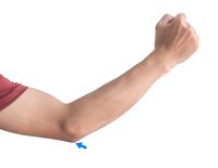
The proximal end of the ulna forms a bony protrusion, called the olecranon, and constitutes the point of the elbow.
joints. The elbow is made up of three joints (1):
- the humero-ulnar joint, connecting the humeral trochlea, in the form of a pulley, and the throchlear notch of the ulna (or ulna). These two surfaces are covered with cartilage;
- the humeral-radial joint connecting the capitulum of the humerus and the radial dimple;
- the proximal radio-ulnar joint connecting the two ends of the radius and ulna laterally.
Insertions. The elbow region is the place of insertions of many muscles and ligaments allowing movement of the elbow and maintaining the structure.
Elbow joint
Elbow movements. The elbow can perform two movements, flexion, which brings the forearm closer to the arm, and extension, which corresponds to the reverse movement. These movements are done mainly through the humero-ulnar joint and to a lesser extent through the humero-radial joint. The latter is involved in the direction of movement and in the amplitude, which can reach 140 ° on average. (2)
Forearm movements. The elbow joints, mainly the radio-ulnar joint and to a lesser extent the humero-radial joint, are involved in the pronosupination movements of the forearm. Pronosupination is made up of two distinct movements (3):
— The supination movement which allows the palm of the hand to be oriented upwards
— The pronation movement which allows the palm of the hand to be oriented downwards
Fracture and pain in the elbow
fractures. The elbow can suffer from fractures, one of the most frequent of which is that of the olecranon, located at the level of the proximal epiphysis of the ulna and forming the point of the elbow. Fractures of the radial head are also common.
osteoporosis. This pathology constitutes a loss of bone density which is generally found in people over the age of 60. It accentuates bone fragility and promotes bills (4).
Tendinopathies. They designate all the pathologies that can occur in the tendons. The symptoms of these pathologies are mainly pain in the tendon during exertion. The causes of these pathologies can be varied. Epicondylitis, also called epicondylalgia, refers to pain occurring in the epicondyle, a region of the elbow (5).
Tendinitis. They refer to tendinopathies associated with inflammation of the tendons.
Treatments
Medical treatment. Depending on the pathology diagnosed, different treatments may be prescribed to regulate or strengthen bone tissue, as well as to reduce pain and inflammation.
Surgical treatment. Depending on the type of fracture, a surgical operation can be carried out with, for example, the installation of a screwed plate, nails or even an external fixator.
Arthroscopy. This surgical technique allows the joints to be observed and operated on.
Physical treatment. Physical therapies, through specific exercise programs, are most often prescribed such as physiotherapy or physiotherapy.
Elbow examination
Physical examination. Diagnosis begins with an assessment of forearm pain to identify its causes.
Medical imaging exam. X-ray, CT, MRI, scintigraphy or bone densitometry examinations can be used to confirm or deepen the diagnosis.
History
External epicondylitis, or epicondylalgia, of the elbow is also referred to as “tennis elbow” or “tennis player’s elbow” since they occur regularly in tennis players. (6) They are much less common today thanks to the lighter weight of current rackets. Less frequent, internal epicondylitis, or epicondylalgia, are attributed to the “golfer’s elbow”.