


Contents
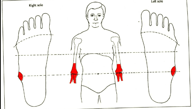
Elbow (Foot)
Ulna anatomy
Position. Elongated bone, the ulna, or ulna, forms the skeleton of the forearm with the radius. The ulna starts at the elbow and extends to the wrist.
Structure. The ulna, or ulna, can be divided into three different elements:
- the central part, called diaphysis, having a triangular shape when cut
- the two ends: the proximal epiphysis articulating with the radius and the humerus to form the elbow; and the distal epiphysis, which joins the radio-ulnar articular disc, at the wrist. (1)
Ulna movements
The ulna, or ulna, allows pronosupination movements of the forearm thanks to the joints at the elbow and wrist. 2 Pronosupination is made up of two distinct movements:
- The supination movement which allows the palm of the hand to be oriented upwards
- The pronation movement which allows the palm of the hand to be oriented downwards
Pathologies of the elbow fracture du
fractures. The forearm, and especially the ulna, is often the site of fractures. (3)
- Fracture of the olecranon. Located at the level of the proximal epiphysis of the ulna and forming the point of the elbow, the olecranon can be impacted and fracture. Frequent in the ulna, this fracture is usually due to a direct impact on the forearm.
Bone pathologies.
- Osteoporosis. This is the loss of bone density which generally affects individuals over the age of 60. It results in bone fragility and a risk of fractures. (4)
- Bone cancer. Metastases can develop in the bones. These cancer cells usually originate from primary cancer in another organ. (5)
- Bone dystrophy. This pathology constitutes an abnormal development or remodeling of bone tissue and includes many diseases. One of the most common, Paget’s disease (6) causes bone densification and deformation, leading to pain. Algodystrophy is the appearance of pain and / or stiffness following a trauma (fracture, surgery, etc.).
Treatment and prevention of ulna
Drug treatments. Depending on the disease, different treatments are prescribed to regulate or strengthen bone tissue or reduce pain and inflammation.
Surgical treatment. Depending on the type of fracture, a surgical operation can be performed with, for example, the placement of pins, a screwed plate or even an external fixator.
Orthopedic treatment. Depending on the type of fracture, the installation of a plaster, a resin can be carried out to immobilize the ulna.
Hormonal treatment, radiotherapy or chemotherapy. These treatments may be prescribed depending on the stage of cancer progression.
Examen du elbow
Medical imaging exam. Depending on the suspected or proven pathology, additional examinations may be performed such as an X-ray, an ultrasound, a CT scan, an MRI or a scintigraphy.
Medical analysis. In order to identify certain pathologies, blood or urine analyzes can be carried out such as, for example, the dosage of phosphorus or calcium.
Bone biopsy. In some cases, a bone sample is taken to confirm a diagnosis.
History and symbolism of the ulna
In 2014, the National Center for Scientific Research (CNRS) as well as the National Institute of Preventive Archeology unveiled the discovery of arm and forearm bones, including that of the ulna, on the banks of the Seine in Normandy. . These bones belonged to a pre-Neanderthal ancestor dating back 200 years. (000)