


Contents
CSF: the role and pathologies associated with cerebrospinal fluid
Cerebrospinal fluid is a fluid that bathes the structures of the central nervous system: the brain and the spinal cord. It has a role of protection and shock absorber. The cerebrospinal fluid is in a normal state, devoid of germs. The appearance of a germ in it can be responsible for serious infectious pathologies.
What is cerebrospinal fluid?
Definition
Cerebrospinal fluid or CSF is a fluid enveloping the central nervous system (brain and spinal cord). It circulates through the ventricular system (ventricles located in the brain) and the subarachnoid space.
As a reminder, the central nervous system is surrounded by envelopes called the meninges, made up of 3 layers:
- the dura, a thick outer layer;
- the arachnoid, a thin layer between the dura and the pia mater;
- the pia mater, internal thin sheet, adhering to the cerebral surface.
The space between the arachnoid and the pia mater corresponds to the subarachnoid space, place of circulation of the cerebrospinal fluid.
Features
The total daily production of CSF is estimated to be approximately 500 ml.
Its volume is 150 – 180 ml, in adults, and so it is renewed several times a day.
Its pressure is measured using a lumbar puncture. It is estimated between 10 and 15 mmHg in adults. (5 to 7 mmHg in infants).
To the naked eye, CSF is a clear liquid said to be rock water.
Composition
Celphalo-spinal fluid is made up of:
- water;
- leukocytes (white blood cells) <5 / mm3;
- of proteins (called proteinorrachia) between 0,20 – 0,40 g / L;
- glucose (known as glycorrachia) represents 60% of glycemia (blood sugar level), or approximately 0,6 g / L;
- many ions (sodium, chlorine, potassium, calcium, bicarbonate)
The CSF is completely sterile, that is to say does not contain pathogenic microorganisms (viruses, bacteria, fungi).
Cerebrospinal fluid: secretion and circulation
Features
Cerebrospinal fluid is a fluid that bathes the structures of the central nervous system. It has a role of protection and shock absorber of the latter, in particular during movements and changes of position. Cerebrospinal fluid is normal, germ free (sterile). The appearance of a germ in it can be responsible for serious infectious pathologies that can lead to neurological sequelae or even the death of the patient.
Secretion and circulation
The cerebrospinal fluid is produced and secreted by the choroid plexuses corresponding to structures located at the level of the walls of the different ventricles (lateral ventricles, 3rd ventricle and 4th ventricle) and making it possible to make a junction between the blood system and the central nervous system .
There is a continuous and free circulation of the CSF at the level of the lateral ventricles, then to the 3rd ventricle through the Monroe holes and then to the 4th ventricle through the Sylvius aqueduct. It then joins the subarachnoid space through the foramina of Luscka and Magendie.
Its reabsorption takes place at the level of the arachnoid villi of Pacchioni (villous growths located on the external surface of the arachnoid), allowing its flow to the venous sinus (more exactly the upper longitudinal venous sinus) and thus its return to the venous circulation. .
Examination and analysis of cerebrospinal fluid
The analysis of the CSF makes it possible to detect many pathologies, most of which require urgent care. This analysis is performed by a lumbar puncture, which consists of taking the CSF, by inserting a thin needle between two lumbar vertebrae (the majority of cases, between the 4th and 5th lumbar vertebrae in order to avoid any risk of damage to the spinal cord. , stopping opposite the 2nd lumbar vertebra). Lumbar puncture is an invasive act, which must be performed by a doctor, using asepsis.
There are contraindications (severe coagulation disorder, signs of intracranial hypertension, infection at the puncture site) and side effects may occur (post-lumbar puncture syndrome, infection, hematoma, lower back pain).
The CSF analysis includes:
- a macroscopic examination (examination with the naked eye allowing the appearance and color of the CSF to be analyzed);
- a bacteriological examination (search for bacteria with the realization of cultures);
- a cytological examination (looking for the number of white and red blood cells);
- a biochemical examination (search for the number of proteins, glucose);
- additional analyzes can be performed for specific viruses (Herpes virus, Cytomegalovirus, Enterovirus).
Cerebrospinal fluid: what associated pathologies?
Infectious pathologies
Meningitis
It corresponds to the inflammation of the meninges which in most cases is secondary to infection by a pathogenic agent (bacterial, virus or even parasite or fungi) due to contamination of the cerebrospinal fluid.
The main symptoms of meningitis are:
- diffuse and intense headaches with discomfort from noise (phonophobia) and light (photophobia);
- a fever ;
- nausea and vomiting.
On clinical examination, one can detect meningeal stiffness, that is to say an invincible and painful resistance when bending the neck.
This is explained by a contraction of the para-vertebral muscles in connection with the irritation of the meninges.
If meningitis is suspected, it is essential to completely undress the patient, in order to look for signs of purpura fulminans (skin hemorrhagic spot linked to a coagulation disorder, which does not disappear when pressure is exerted). Purpura fulminans is a sign of a very severe infection, most often secondary to infection with meningococcus (bacteria). It is a life-threatening emergency requiring an intramuscular or intravenous injection of antibiotic therapy as quickly as possible.
Additional examinations are often necessary for the certainty of the diagnosis:
- lumbar puncture (except in cases of contraindication) allowing an analysis to be carried out;
- biological assessment (blood count, hemostasis assessment, CRP, blood ionogram, glycemia, serum creatinine, and blood cultures);
- urgent brain imaging in the following cases that contraindicate lumbar puncture: disturbance of consciousness, neurological deficit and / or seizure.
The analysis of the CSF makes it possible to direct towards a type of meningitis and to confirm the presence of a pathogenic agent.
Treatment will depend on the type of germ present in the cerebrospinal fluid.
Meningoencephalitis
It is defined by the association of an inflammation of the brain and the meningeal envelopes.
It is based on the association of a meningeal syndrome (headache, vomiting, nausea and meningeal stiffness) and an impairment of the brain directed by the presence of disorders of consciousness, partial or total convulsive seizures or even sign of a neurological deficit (motor deficit, aphasia).
Meningoencephalitis is a serious pathology that can lead to the death of the patient and therefore requires urgent medical care.
A suspicion of meningoencephalitis requires urgent brain imaging, and must be performed before the lumbar puncture.
Other additional examinations confirm the diagnosis:
- a biological assessment (blood count, CRP, blood ionogram, blood cultures, hemostasis assessment, serum creatinine);
- an EEG (electroencephalogram) may be performed, which may show signs in favor of brain damage.
The management by a medical treatment must be rapid and will then be adapted to the revealed germ.
Carcinomatous meningitis
Carcinomatous meningitis is inflammation of the meninges due to the presence of cancer cells found in the CSF. More exactly, it is a question of metastases, that is to say a secondary dissemination resulting from a primary cancer (in particular from lung cancer, melanoma and breast cancer).
The symptoms are polymorphic, consisting of:
- meningeal syndrome (headache, nausea, vomiting, stiff neck);
- disturbances of consciousness;
- behavioral change (memory loss);
- seizures;
- neurological deficit.
Additional examinations are necessary to confirm the diagnosis:
- performing a brain imaging (brain MRI) which can show signs in favor of the diagnosis;
- a lumbar puncture to look for the presence of cancer cells in the CSF and thus confirm the diagnosis.
The prognosis of carcinomatous meningitis is still gloomy today with few effective therapeutic means.
Hydrocephalus
Hydrocephalus is an accumulation of an excessive amount of cerebrospinal fluid within the cerebral ventricular system. It is demonstrated by performing a brain imaging which finds dilation of the cerebral ventricles.
This excess can result in an increase in intracranial pressure. Indeed, the intracranial pressure will depend on several parameters which are:
- the brain parenchyma;
- cerebrospinal fluid;
- cerebrovascular volume.
So when one or more of these parameters are modified, it will have an impact on the intracranial pressure. Intracranial hypertension (HTIC) is defined as a value> 20 mmHg in adults.
There are different types of hydrocephalus:
- non-communicating hydrocephalus (obstructive): it corresponds to an excess accumulation of cerebrospinal fluid in the ventricular system secondary to an obstacle affecting the circulation of the CSF and thus to its reabsorption. Most often, it is due to the presence of a tumor compressing the ventricular system, but also can be secondary to malformations present from birth. It results in an increase in intracranial pressure requiring urgent treatment. It is possible to carry out an external ventricular bypass of the CSF (temporary solution) or even more recently developed, the realization of an endoscopic ventriculocisternostomy (creation of a communication between the cerebral ventricular system and the cisterns which correspond to an enlargement of the subarachnoid space) thus allowing to bypass the obstacle and to find an adequate flow of the CSF;
- communicating hydrocephalus (non-obstructive): it corresponds to an excess accumulation of cerebrospinal fluid in connection with a gene in the reabsorption of CSF. It is most often secondary to subarachnoid hemorrhage, head trauma, meningitis or possibly idiopathic. It requires management by an internal CSF shunt called ventriculoperitoneal shunt (if the fluid is directed to the peritoneal cavity) or ventriculo-atrial shunt (if the fluid is directed to the heart);
- chronic hydrocephalus at normal pressure: it corresponds to an excess of cerebrospinal fluid in the cerebral ventricular system but without increase in intracranial pressure. It most often affects adults, after 60 years with a predominance of men. The pathophysiological mechanism is still poorly understood. It can be found in people with a history of subarachnoid hemorrhage, head trauma or having had intracranial surgery.
It is defined most of the time by a triad of symptoms, called the Adams and Hakim triad:
- memory impairment;
- sphincter disorders (urinary incontinence);
- trouble walking with slow walking.
Brain imaging can show a dilation of the cerebral ventricles.
Management is based mainly on the establishment of an internal ventricular bypass, either ventriculo-peritoneal or ventriculo-atial.
Other pathologies
Analysis of cerebrospinal fluid can reveal many other pathologies:
- subarachnoid hemorrhage with evidence of blood circulating in the CSF;
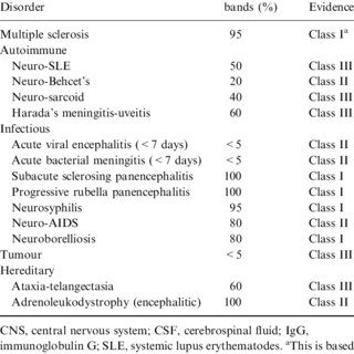
- inflammatory diseases affecting the central nervous system (multiple sclerosis, sarcoidosis, etc.);
- neurodegenerative diseases (Alzheimer’s disease);
- neuropathies (Guillain-Barré syndrome).