


Carotid
The carotids are arteries supplying the brain, neck and face. Carotid stenosis is the main pathology to be feared. Relatively common with age, it may or may not lead to a transient stroke.
Anatomy
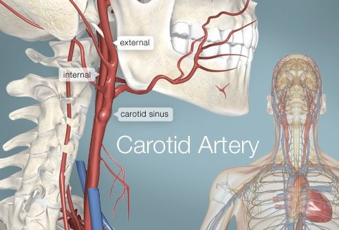
The brain is supplied by different arteries: two carotid arteries in front and two vertebral arteries behind. These four arteries meet at the base of the skull to form what is called the Polygon of Willis.
The so-called primary or common carotid artery arises from the aorta and ascends in the neck. It divides at the level of the middle part of the neck into two arteries: the internal carotid and the external carotid. This junction zone is called carotid bifurcation.
physiology
The internal carotid arteries supply to the brain, while the external carotid arteries supply to the neck and face. These are therefore very important arteries.
Anomalies / Pathologies
Carotid stenosis is the main lesion to fear in the carotid artery.
It corresponds to a decrease in the diameter of the carotid artery, most often following the formation of an atheromatous plaque (deposition of cholesterol, fibrous and calcareous tissues) within the artery. In the majority of cases (90%), this stenosis is localized at the level of the cervical carotid bifurcation.
The risk is that the carotid artery will end up being blocked by the atheromatous plaque or that it will fragment. A transient ischemic attack (TIA) can then occur which regresses without sequelae in less than 24 hours, or a cerebrovascular accident (AVC) or cerebral infarction, with more or less serious sequelae.
Carotid stenosis is common with age: according to the Haute Autorité de Santé, 5 to 10% of people over 65 have a stenosis greater than 50%. Carotid stenosis is estimated to be responsible for about a quarter of strokes.
Treatments
The management of carotid stenosis is based on drug treatment, control of vascular risk factors and for some patients a revascularization procedure.
Regarding drug treatment, three types of drugs are prescribed together: an antiplatelet agent to thin the blood, a statin to limit the development of atheromatous plaques and an ACE inhibitor (or beta blocker in some cases).
Regarding revascularization, the French National Authority for Health has issued specific recommendations for the indication of surgery according to the degree of symptomatic carotid stenosis:
- between 70 and 99% of stenosis, surgery is indicated with an equivalent significant benefit in men and women;
- between 50 and 69% stenosis, surgery may be indicated but the benefit is less, especially in women;
- between 30 and 49%, surgery is not useful;
- below 30%, the surgery is deleterious and should not be performed.
When revascularization is indicated, surgery remains the gold standard. The procedure, called carotid endarterectomy, is most often performed under general anesthesia. The surgeon makes an incision in the neck, clamps the three arteries and then cuts the carotid artery at the level of the stenosis. He then carefully removes the atherosclerotic plaque and its debris, then closes the artery with a very fine wire.
Angioplasty with a stent is not indicated as a first-line treatment. It is only offered in certain specific cases of contraindication to surgery.
In case of asymptomatic carotid stenosis:
- greater than 60%: revascularization by carotid surgery may be indicated depending on certain factors (life expectancy, progression of the stenosis, etc.);
- in case of stenosis less than 60%, surgery is not indicated.
Along with drug and surgical treatment, it is essential to review your lifestyle to limit the risk factors: high blood pressure, tobacco, hypercholesterolemia and diabetes.
Diagnostic
Carotid stenosis can be asymptomatic and be discovered during a medical examination by your general practitioner or specialist, or during an ultrasound of the thyroid for example. The presence of a carotid murmur on auscultation should lead to the prescription of a carotid doppler ultrasound to diagnose a possible carotid stenosis and assess the rate of obstruction. Depending on the results, MRI angiography, CT angiography or digital carotid angiography will be prescribed. It makes it possible to determine the location, morphology and extension of the plaque, and to assess the diffusion of the atheroma on the other axes and in particular the other carotid artery.
When symptomatic, the signs of carotid stenosis are those of transient ischemic attack (TIA) and stroke. Either, depending on the area of the brain affected:
- eye damage (sudden and painless loss of vision in one eye or transient amaurosis);
- paralysis on one side of the body, either total or limited to the upper limb and / or the face (hemiparesis, facial paralysis);
- loss of speech (aphasia).
Faced with these signs, it is essential to contact 15.