


Contents
The varicose wound
A wound in the leg that does not heal? It may be a varicose wound, in other words a varicose ulcer. It occurs at the last stage of the evolution of chronic venous insufficiency secondary to varicose veins or to sequelae of phlebitis. Even if it is not very painful, it requires appropriate local treatments, accompanied by management of the venous disease in question to avoid recurrence.
What is a varicose sore?
Definition
Varicose veins, otherwise known as varicose ulcers or venous ulcers, are a complication of varicose veins or phlebitis that usually occurs after a long period of development.
It presents as a wound in the leg – classically in the ankle – with loss of skin substance, the healing time of which is greater than one month. Left untreated, it can become superinfected and persist for months or even years.
A venous ulcer is distinguished from an arterial ulcer, which results from arterial disease of the lower limbs, usually linked to atherosclerosis or diabetes.
Causes
The varicose wound occurs in the late phase of evolution of chronic venous insufficiency. The superficial or deep veins no longer provide a correct venous return to the heart and the blood tends to stagnate.
- In people with varicose veins, there is a loss of elasticity of the veins as well as a dysfunction of the valves equipping the wall of the vessels, whose role is to prevent reflux.
- Venous insufficiency can also be due to the sequelae of phlebitis (venous thrombosis). In this case, the stagnation of the blood and the increase in blood pressure eventually lead to irreversible damage to the valves.
- More rarely, a congenital disease, primary deep valve insufficiency, is responsible for venous insufficiency.
- A deficiency of the calf muscle pump is also often found.
In all cases, stasis (blood stagnation) causes hypertension in the legs and ankles as well as the leakage of inflammatory fluid. Tissue suffering is linked to the presence of toxins and the lack of nutrient and oxygen supply. It results in their destruction (necrosis).
Diagnostic
The clinical examination carried out by the phlebologist makes it possible to make the diagnosis and assess the severity of the wound. Measurements and photos of the wound can be taken.
Knowledge of the patient’s history (phlebitis, age of varicose veins, etc.) is useful.
The doctor also seeks to ensure that arterial damage is not involved in the origin of the ulcer. He will be able to look for associated symptoms (in particular pain and intermittent claudication), to feel the arterial pulses and to measure the pressure at the level of the ankle.
Venous echo-doppler
This imaging test is used to visualize blood flow and assess its speed. It is used to identify the origin of the varicose ulcer.
Additional tests
Various examinations make it possible to refine the diagnosis:
- blood tests,
- bacterial samples,
- biopsies…
The people concerned
The frequency of venous ulcer increases with age. In some studies, leg ulcers (linked 9 times out of 10 to venous damage), affect up to 1% of the general population, 3% of over 65s and 5% of over 80s.
There is a clear female predominance of the disease.
Risk factors
These are those of venous insufficiency:
- heredity,
- in women, hormonal status,
- prolonged standing posture,
- physical inactivity,
- overweight,
- smoking,
- repeated exposure to heat (very hot baths, underfloor heating, etc.) …
Symptoms of varicose sore
Warning signs
Chronic venous insufficiency is manifested by various symptoms: heavy legs, edema, presence of spider veins (small purplish venules on the surface) or varicose veins, cramps, etc.
Skin changes usually precede the formation of the varicose wound:
- ocher dermatitis (ocher skin spots),
- a whitish atrophy,
- hypodermatitis (inflammation of the deep dermis),
- varicose eczema (reddish itchy patches).
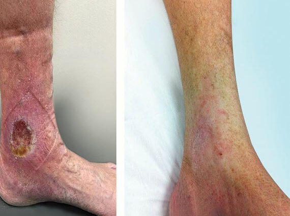
Evolution of the lesion
The varicose wound sits below the knee, usually at the ankle, in the area of the malleolus. It can appear as a result of intense scratching or a minor shock.
The skin cracks and forms a crater with irregular and reddish edges, sometimes very spectacular in appearance.
The appearance of the wound varies depending on the stage of development:
- Tissue necrosis is first indicated by a blackish color.
- In the fibrous stage, the wound becomes covered with a yellowish coating and oozes frequently. The risks of infection are high. Purulent wounds have a greenish appearance.
- The healing process is difficult. It first results in fleshy buds, before the epidermis comes to cover the wound.
It should also be noted that an arterial ulcer sits more frequently in the foot, in areas of friction.
pain
Varicose wounds are often not very painful. Significant pain suggests the presence of an arterial component or superinfection.
Treatment of varicose wounds
Local care
Performed by a nurse, the local care must be adapted to the stage of evolution of the ulcer. Healing requires regular care (several times a week) over fairly long periods.
The wound is first carefully cleaned, conventionally with soap and water or using a betadine-type solution when the wound is infected. If necessary, the nurse performs a debris, that is to say a deep cleaning with removal of fibrinous debris.
The care is completed by the break of a suitable dressing, for example:
- fatty dressings if the wound is dry,
- absorbent dressings (hydrocellular, alginates) in the event of exudation,
- hemostatic dressings (alginates) in case of blood flow,
- silver dressings in case of superinfection.
Honey dressings have been tried in the treatment of venous ulcers, but do not appear to be effective.
Compression (venous retention)
Treatment of the cause of the varicose wound is imperative. Elastic compression is used to reduce local edema and improve venous return. The doctor adapts his prescription according to the stage of healing of the wound, the presence or absence of edema and the patient’s tolerance.
Different devices exist, which must be worn either 24 hours a day, or from sunrise to sunset:
- Multilayer bandages (several superimposed bands) are generally the most suitable at the start of treatment,
- simple elastic bands or elastic compression stockings are often offered as a second step.
Treatment of varicose veins
Usually necessary to prevent recurrence, the treatment of varicose veins involves, in particular, sclerotherapy and venous surgery.
The transplant
Skin grafts in pastilles or mesh are possible when a varicose ulcer resists conventional treatments for more than 6 months.
Global support
The doctor ensures that the anti-tetanus vaccination is up to date. The management can also include hygieno-dietetic measures (fight against overweight or against undernutrition), pain relief treatment, lymphatic drainage performed by a physiotherapist, etc.
Prevent varicose wounds
The prevention of varicose wounds is based on the same principles as that of venous insufficiency.
The rules of hygiene of life play an essential role. Physical activity stimulates blood circulation and prevents the appearance of varicose veins. We recommend that you walk at least 30 minutes a day, at least three times a week. More specifically, all sports that work the calves (cycling, dancing, etc.) improve venous return.
Other measures (sleeping with raised feet, avoiding too hot baths, sauna, underfloor heating, prolonged exposure to the sun or even tight clothing that impedes blood circulation, etc.) are especially necessary in people with already poor circulation. Also watch out for air travel!
We will also preserve our venous capital by keeping a healthy weight, adopting a balanced diet and avoiding smoking.