


The embryo: the development of the embryo during pregnancy
During the first 8 weeks of pregnancy, the future baby evolves at high speed … Cell division, formation of its organs and its appendages, the embryo then goes through the period known as embryogenesis. What are the major first stages of intrauterine life? Decryption.
Definition of the embryo
We speak of an embryo from the appearance of the first cell following the fusion between the spermatozoon and the oocyte. The embryonic phase then corresponds to the growth and development of the unborn child from this very first stage until the 8th week of pregnancy (10 weeks), i.e. 56 days after fertilization.
Described in medicine by the 23 stages of Carnegie, this key period of intrauterine life can be more simply divided into 2 main phases:
- the formation and delimitation of the embryo from fertilization to the 4th week of pregnancy,
- the outline of the embryonic organs, until the 8th week of pregnancy.
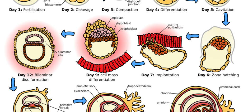
The development of the embryo: from the zygote to the blastocyst
Following fertilization, embryogenesis begins with the zygote, a single cell born from the fusion of male and female gametes and already carrying the genetic information of the future baby. In the hours following its formation, the zygote begins to divide, by a phenomenon of mitosis, into 2 cells of equal size (the blastomeres), then into 4, then into 8 around the 60th hour after fertilization, etc. This is the so-called stage of the segmentation.
Between 72 hours after fertilization and the 4th day of pregnancy, the embryo begins his migration from the fallopian tube to the uterus while cell division continues. Then composed of 16 cells, the embryo resembles a blackberry, hence its name morula. The morula then evolves into a blastocyst, a stage at which the cells differentiate:
- the peripheral cell layer, the trophoblast, is at the origin of the embryonic appendages which will later constitute the placenta,
- the 3 or 4 most central (and bulky) cells of the blastocyst form an internal cell mass from which the embryo will evolve: it is the embryoblast or embryonic button.
Between the 4th and 5th day after fertilization, the embryo finishes its journey in the uterine cavity. It then loses its protective envelope, the zona pellucida. Also called hatching, this key step facilitates the attachment of the embryo to the uterine lining, and finally 7 days after fertilization, implantation.
Embryonic phase: the primitive layers of the embryo
During the second and third week of pregnancy (4 and 5 weeks), the cluster of cells which until then constituted the embryo evolves into an embryonic disc composed of 2 then 3 layers (or primitive layers). We then speak of gastrulation. From these sheets will result the tissues and organs of the unborn child and more particularly:
- of the ectoblast, external layer, will be born part of the nervous system, the epidermis, the mucous membranes or the teeth.
- from l’endoblaste, internal layer, will result the organs of the digestive and respiratory system as well as the liver and the pancreas in particular.
- du mesoblast will appear somites (at the origin of muscles, ligaments, skin or even cartilage.), gonads (future sex cells), kidneys or the circulatory system.
Development of the embryo: the delineation of the embryo
Embryogenesis passes a new key stage during the 4th week of pregnancy (6 weeks). The primitive layers then evolve into a cylindrical C-shaped structure, under the effect of the folding of the embryonic disc. This delimitation of the embryo, a phenomenon allowing its circumscription in relation to the appendages and thus prefiguring its future anatomy, takes place in 2 stages:
- When bending in the transverse direction, the future back of the embryo, at this stage described as the dorsal protrusion, appears, the volume of the amniotic cavity increases, the embryo and its appendages fold back on themselves.
- During longitudinal inflection, the cranial and caudal regions of the embryo come together
Well defined, now floating in the amniotic cavity, the embryo continues to develop:
buds of the upper limbs appear, the heart begins to beat, the first 4-12 somites are visible on its dorsal side.
The embryonic phase and organogenesis
From the second month of pregnancy, the organs of the embryo are developing at high speed. It is organogenesis.
- Under the effect of the rapid development of the nervous system, the cephalic pole of the embryo (its head) grows and flexes. Inside, the forebrain (forebrain) divides in two around the 5th week of pregnancy. Another notable phenomenon at this stage: the outline of the sense organs.
- Around the 6th week, it is at the beginnings of the external auditory canal to appear, just like the vertebrae, currently placed around the spinal cord, and the back muscles. Other characteristics of the embryo at this stage: its stomach has its final shape and the primitive sex cells are in place.
- At 7 weeks pregnant, the limbs continue to grow and the inter-digital grooves appear on the hands and toes while the musculature of the heart becomes different.
By the end of the 8th week, organogenesis is almost complete. The organs are differentiated and will only have to “grow” during the fetal phase. The embryo, for its part, takes on an increasingly human form: its head stands up, its neck is now formed just like its face and more particularly its lips, nose, eyes and ears.
When the embryo becomes a fetus
At 9 weeks of pregnancy (11 weeks), the embryo becomes a fetus. The fetal period, which lasts from the 3rd month of gestation until childbirth is characterized above all by the growth of tissues and organs. It is also during this phase that the fetus experiences a significant increase in size and weight. A particularly telling example: from 3 cm and 11 g at the end of the embryonic period, the future baby passes to 12 cm and 65 g at the end of the 3rd month of pregnancy!