


Contents
Stomach
The stomach (from the Latin stomachus meaning “esophagus” and by extension “stomach”) is an organ of the digestive tract. Food storage area, it is at this level that the digestion process begins.
Stomach anatomy
The stomach is a sac-like organ on the left side of the abdominal cavity, partly hidden by the liver and diaphragm. Its dimensions vary depending on the individual and the meal, but on average it is 25 cm long, 10-15 cm wide and can hold up to 4L of food.
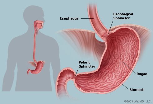
It is part of the digestive tract: it is connected to its entrance to the esophagus and its exit to the small intestine.
It is divided into 5 regions:
- The cardia is the area in the upper part of the stomach, near the esophagus. Food enters the stomach through the orifice of the cardia. At its level is the cardial sphincter muscle which prevents gastric reflux to the esophagus.
- The fundus is the dome-shaped region above the cardia. Food is temporarily stored at this level.
- The body of the stomach is the main area of the organ, it is at this level that the breakdown of food takes place.
- The pyloric lair follows the body down the stomach. Narrowed area, it stores decomposed food while waiting for it to be discharged into the intestine.
- The pylorus is the last part of the stomach that connects with the small intestine. Funnel-shaped, it is closed by the pyloric sphincter muscle which controls the emptying of stomach contents into the small intestine.
- The small curvature, on the concave medial face,
- The large curvature corresponds to the convex side face.
Two main curvatures define the shape of the stomach:
- The small curvature, on the concave medial face,
- The large curvature corresponds to the convex side face.
Functions of the stomach
The stomach temporarily stores food. But it is also the seat of the breakdown of solid foods into a kind of porridge called “chyme”.
When food arrives in the stomach, it is broken into fragments. This step is made possible by the contraction of the stomach muscle, the muscularis. Composed of three layers of fibers arranged in a longitudinal, circular and oblique manner, it mixes and kneads the food.
Then begins the chemical digestion. Only the digestion of proteins is initiated in the stomach. The mucous membrane contains glands which secrete gastric juices. Under normal conditions, the production of juices is 2 to 3 L per day. Hydrochloric acid and enzymes are also produced by cells in the stomach. All of these substances are mixed with food and break it down. Crushed, it is transformed into liquid, the chyme. At this stage, the chyme is at the level of the pylorus. It is then gradually propelled into the small intestine by contraction of the stomach. Usually, the stomach empties in about 4 hours after a balanced meal and a minimum of 6 hours for a high fat meal.
Interestingly, a mucus coats the lining of the stomach to counter the corrosive effects of hydrochloric acid and enzymes.
Stomach pathologies
aerophagia : physiological phenomenon characterized by abnormally high air ingestion during swallowing. Air collects in the esophagus and sometimes a small amount in the stomach when the subject drinks or eats, causing bloating and belching (burping).
Gastroesophageal reflux disease (or heartburn) : refers to the ascent of part of the contents of the stomach into the esophagus. This content is very acidic and the lining of the esophagus is not designed to withstand such acidity, which causes it to become inflamed which results in sensations of burning or irritation.
Stomach cancer : develops from a parietal cell (cell in the wall of the stomach), initially normal, which multiplies in an anarchic fashion to form a mass called a malignant tumor. It is a cancer that progresses slowly and that is rarely seen before the age of 50.
Gastroenteritis : infection of the digestive system which causes nausea, vomiting, abdominal cramps and diarrhea. In the majority of cases, it is short-lived, the symptoms appear quickly and usually disappear after 1 to 3 days. It is most often caused by a virus or bacteria that are transmitted mainly through contaminated hands, water and food (food poisoning).
Ulcer : inflammation of the stomach wall. The ulcer is linked to the proliferation of bacteria in the stomach, Helicobacter pylori, qui invades the mucus layer that normally protects the stomach from acidity. It then disrupts the protective mechanism in some people. This layer of mucus disappears and then it is the ulcer itself which causes damage to the inner lining of the stomach.
Gastritis : inflammation of the stomach lining. Results in heartburn or difficulty digesting. Usually not serious, gastritis can be caused by alcohol, medication or even l’Helicobacter pylori.
Hernie hiatale : it is the stomach which goes up in part through a small opening called “oesophageal hiatus”, located in the diaphragm, the respiratory muscle which separates the thoracic cavity from the abdomen.
Treatments and prevention of the stomach
Smoking and obesity strongly contribute to the onset of gastroesophageal reflux disease. No convincing prevention exists, but certain measures can reduce burns. Watching your diet is key. Avoiding eating foods that are too fatty, for example, can help because fat slows down the evacuation of food from the stomach and reflux is more important when the stomach is full.
Several preventive measures exist to prevent the spread of gastroenteritis. This disease spreads very easily and knowing that 80% of germs are transmitted through the hands (3), a good wash is your main ally against contamination. The World Health Organization (WHO) also considers that this is the most important hygiene measure to prevent the transmission of these infections.
Taking certain medications can cause stomach upset. This is particularly the case with nonsteroidal anti-inflammatory drugs (NSAIDs) such as aspirin or ibuprofen widely used against rheumatic diseases. These substances increase the risk of gastric complications. In some cases, a medicine that protects the stomach may be combined with taking NSAIDs. However, this prescription should not be systematic, precise Health Insurance (4) in a 2009 report, “this decision is based, for each patient, on his age or the analysis of his risk factors for gastroduodenal complication”. The National Agency for the Safety of Medicines and Health Products (ANSM) (5) advises “to use NSAIDs at the minimum effective dose, for the shortest possible duration”.
Stomach exams
Fibroscopie digestive « haute » : reference examination which makes it possible to visualize the inside of the upper digestive tract (esophagus, stomach, duodenum) thanks to the introduction of a flexible tube called a fiberscope or endoscope which is equipped with small cameras. We can also talk about a gastroscope. The terms endoscopy and gastroscopy then refer to the same examination. Allows to highlight a gastroesophageal reflux, a hiatus hernia or an ulcer for example.
Ultrasound endoscopy : examination which combines endoscopy (visualization of the inside of the digestive tract) with ultrasound (ultrasound analysis of the wall and organs located beyond the wall). Ultrasound endoscopy is the most powerful examination to study the lining of the esophagus, stomach or duodenum and neighboring organs. It makes it possible to search for and analyze benign or malignant tumors.
X-ray : medical imaging technique using X-rays. To explore the upper digestive tract (esophagus, stomach, duodenum), the examination is preceded by the ingestion of a so-called “contrast” liquid. It helps bring out the digestive tract when taking x-rays.
pHmétrie oesophagienne : corresponds to the measurement of the acidity (pH) of the esophagus. This test is used to diagnose and assess the extent of gastroesophageal reflux disease.
Esophageal manometry: test that measures esophageal contraction and lower sphincter tone. It allows to highlight the existence of such as gastroesophageal reflux.
Anecdote and stomach
Australian researchers Robin Warren and Barry Marshall showed in 1982 that stomach ulcers were caused by the bacteria Helicobacter pylori and can be treated with antibiotics. But in the 1980s, the scientific community believed that ulcers were related to stress and lifestyle. Hence a certain skepticism about this new discovery. To prove their hypothesis, Barry Marshall then infected himself with the bacteria and then treated himself with antibiotics! Warren and Marshall received the 2005 Nobel Prize in Medicine and Physiology for their work on stomach ulcer (9).