


Contents
Pupil
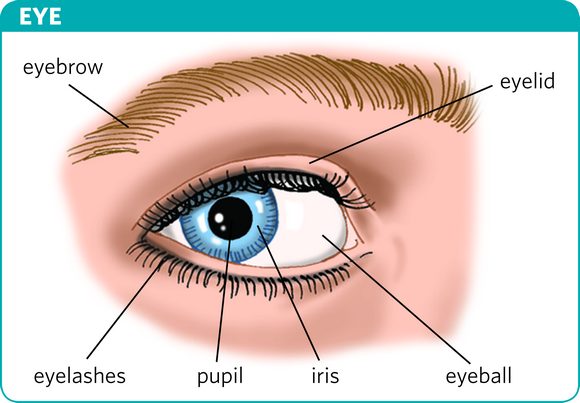
The pupil (from Latin pupilla) is the black circular orifice, located at eye level in the center of the iris.
Anatomy of the pupil
Position. The pupil is the central circular opening of the iris, and allows light to enter the eye. At the level of the eyeball, the pupil and the iris are located between the lens, at the back, and the cornea, at the front. (1)
Structure. The iris is made up of layers of muscle cells that form two muscles (1):
- the sphincter muscle of the pupil, the contraction of which decreases the diameter of the pupil. It is innervated by parasympathetic nerve fibers, participating in the autonomic nervous system.
- the dilator muscle of the pupil, the contraction of which increases the diameter of the pupil. It is innervated by sympathetic nerve fibers, participating in the autonomic nervous system.
Mydriasis
Myosis/Mydriase. Miosis is the narrowing of the pupil while mydriasis is the dilation of the pupil.
Dosage of the amount of light. The iris muscles are used to measure the entry of light into the eye (1):
- Light entry is reduced when the sphincter muscle of the pupil contracts. This is particularly the case when the eye is facing too much light or is staring at a nearby object.
- Light input is increased when the dilator muscle of the pupil contracts. This is especially the case when the eye is facing a weak light input or looking at a distant object.
Pathologies of the pupil
Cataract. This pathology corresponds to an alteration of the lens, located at the back of the pupil. It manifests as a loss of sight, which can lead to blindness. The alteration of the lens is visible by a change in the color of the pupil, which becomes clear or white instead of black.
Adie’s pupil. This pathology, the cause of which is still unknown, results in an alteration of the parasympathetic innervation of the pupil. (2)
Claude Bernard-Horner syndrome. This pathology corresponds to a failure of the sympathetic innervation and the appendages of the eye. The causes of this syndrome can be damage to the nervous system in the midbrain, spinal cord or dissection of a carotid artery. (2)
Oculomotor nerve palsy. The third cranial nerve, nerve III, or oculomotor nerve is responsible for the innervation of a large number of ocular and extraocular muscles including in particular the parasympathetic innervation of the sphincter muscle of the pupil. Paralysis of this nerve can affect vision. (2)
Glaucoma. This eye disease is caused by damage to the optic nerve. It can affect vision.
Presbyopia. Linked to age, it corresponds to a progressive loss of the eye’s capacity to accommodate. It is due to a loss of elasticity of the lens.
Pupil treatments
Pharmacological treatment. Depending on the pathology, different treatments may be prescribed, including eye drops (eye drops). (3)
Symptomatic treatment. For certain pathologies, the wearing of glasses, in particular tinted glasses, may be prescribed. (4)
Surgical treatment. Depending on the type of pathology, a surgical operation may be carried out such as, for example, the extraction of the lens and the implantation of an artificial lens in certain cases of cataracts.
Examinations of the pupil
Physical examination. The examination of pupillary function is systematically carried out during an ophthalmological evaluation (eg: fundus). It allows a lot of information to be provided.
Pharmacological examination. Pharmacological tests with in particular apraclonidine, or even pilocarpine can be carried out to detect an alteration in the pupillary reaction. (3)
Medical imaging examination. MRI, magnetic resonance angiography, computed tomography or even pupillography can be used to complete the diagnosis.
History and symbolism of the pupil
The appearance of red eyes in a photograph is related to the choroid, one of the membranes of the eye bulb, which is rich in blood vessels. When a photograph is taken, the flash may suddenly light up the eyes. The pupil therefore does not have time to retract and lets appear the red choroid. (1)