


Contents
Orthopantomograms
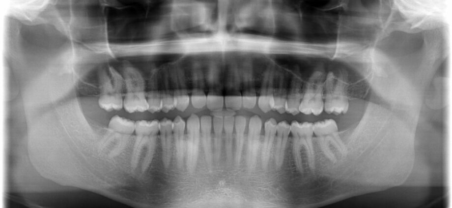
An orthopantomogram is a large dental x-ray, also called a “dental panoramic”, commonly used by dentists. This examination is carried out in a doctor’s office. It is perfectly painless.
What is an orthopantomogram?
An orthopantomogram – or dental panoramic – is a radiology procedure that allows to obtain a very large image of the dentition: the two rows of teeth, the bones of the upper and lower jaw, as well as the jawbone and the mandible. .
More precise and complete than the clinical dental examination, an orthopantomogram makes it possible to highlight lesions of the teeth or gums, invisible or barely visible to the naked eye, such as the beginnings of cavities, cysts, tumors or abscesses. . The dental panoramic also highlights abnormalities of wisdom teeth or impacted teeth.
Dental radiography is also used to know the position of the teeth and their evolution, especially in children.
Finally, it makes it possible to monitor bone loss and the condition of the gums.
All this information is useful for the healthcare practitioner to establish or confirm a diagnosis and define the procedure to follow.
Course of the exam
Prepare for the exam
No special precautions need to be taken before the exam.
Dental appliances, hearing aids, jewelry or bars should be removed just before the examination.
This examination is not possible in a child under two years of age.
During the exam
The dental panoramic takes place in a radiology room.
Standing or seated, you must remain perfectly still.
The patient bites a small plastic support so that the incisors of the upper row and the incisors of the lower row are well placed on the support and the head remains stationary.
When taking the snapshot, a camera moves slowly in front of the face all around the jawbone to scan all of the bones and tissues in the lower face.
The time of the x-ray takes about 20 seconds.
Radiation risks
The radiations emitted by a dental panoramic are far below the maximum authorized dose, and are therefore without risk to health.
Exception for pregnant women
Although the risks are almost zero, all precautions must be taken so that a fetus is not exposed to X-rays. Also, in the event of pregnancy, the doctor must be notified. The latter may then decide to take measures such as protecting the abdomen with a protective lead apron.
Why do a dental panoramic?
There are many reasons for using a dental panoramic. In any case, talk to your dentist.
The health care practitioner may order this examination if he suspects:
- a broken bone
- an infection
- an abscess
- gum disease
- cyst
- a tumor
- bone disease (Paget’s disease for example)
The exam is also useful in monitoring the progress of the ailments mentioned above.
In children, the examination is recommended to visualize the “germs” of future adult teeth and thus assess dental age.
Finally, the doctor will use this x-ray before placing a dental implant to confirm that it is the best option and to determine the location of the roots.
Analysis of the results
A first reading of the results can be carried out by the radiologist or the practitioner carrying out the X-ray. The final results are sent to the doctor or dentist.
Writing : Lucie Rondou, science journalist, December 2018 |
References
- https://www.vulgaris-medical.com/encyclopedie-medicale/panoramique-dentaire/examen-medical
- http://imageriemedicale.fr/examens/imagerie-dentaire/panoramique-dentaire/
- https://www.vulgaris-medical.com/encyclopedie-medicale/panoramique-dentaire/symptomes
- https://www.concilio.com/bilan-de-sante-examens-imagerie-panoramique-dentaire