


Contents
Femoral nerve
The femoral nerve, or crural nerve, provides innervation to different parts of the thigh, hip, and knee.
Femoral nerve: anatomy
Position. The femoral nerve is located in the abdomen and lower limb.
Structure. The femoral nerve is the largest nerve that originates from the lumbar plexus. It is made up of sensory and motor nerve fibers originating from the lumbar vertebrae of the spinal cord, L2 to L4 (1).
Origin. The femoral nerve originates in the abdomen, at the level of the psoas major muscle (1).
Path. The femoral nerve extends and descends posteriorly and laterally to the level of the pelvic girdle.
Branches. The femoral nerve divides into several branches (2):
- The motor branches are intended for the muscles of the anterior part of the thigh, as well as the hip and knee joints (1).
- The sensitive or cutaneous branches are intended for the skin of the front and medial face of the thigh, as well as the medial face of the leg, knee and foot.
Terminations. The terminations of the femoral nerve are (2):
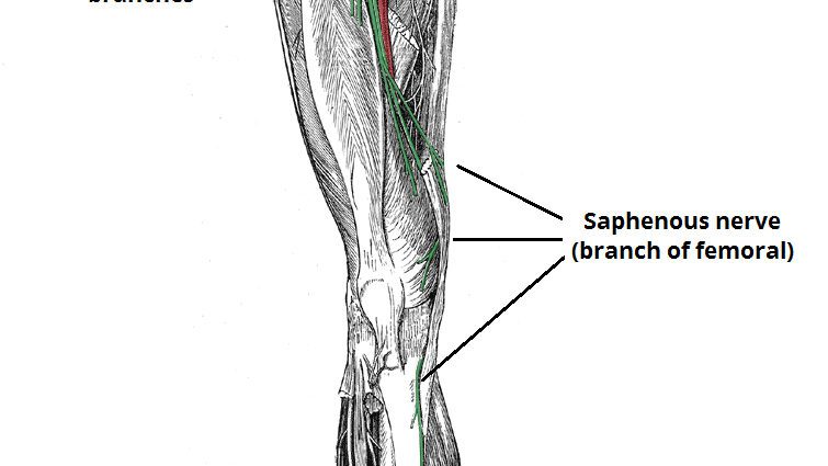
- The saphenous nerve which innervates the medial skin aspect of the leg, foot and hip, as well as the knee joint.
- The medial femoral skin nerve that innervates the anterior and medial skin surfaces of the thigh
- The motor nerve of the thigh muscles that innervates the pectineal, iliac, sartorius, and femoral quadriceps muscles.
Functions of the femoral nerve
Transmission sensitive. The sensitive branches of the femoral nerve make it possible to transmit the different perceptions felt in the skin to the spinal cord.
Drive transmission. The motor branches of the femoral nerve act on the thigh flexor and knee extensor muscles (2).
Degenerative pathologies of the femoral nerve
The various problems associated with the femoral nerve are referred to as cruralgia. These can be manifested by severe pain in the thighs, knees, legs and feet. Their causes are varied but can in particular be of degenerative origin.
Degenerative pathologies. Different pathologies can lead to the progressive degradation of cellular elements. Osteoarthritis is characterized by wear of the cartilage protecting the bones of the joints. (3) The herniated disc corresponds to the expulsion behind the nucleus of the intervertebral disc, by wear of the latter. This can result in the nerves in the spinal cord compressing and reaching the femoral nerve (4).
Treatments
Drug treatments. Depending on the pathology diagnosed, different treatments may be prescribed to reduce pain and inflammation.
Surgical treatment. Depending on the type of pathology diagnosed, surgery may be performed.
- Arthroscopy. This surgical technique allows the joints to be observed and operated on.
Physical treatment. Physical therapies, through specific exercise programs, can be prescribed such as physiotherapy or physiotherapy.
Femoral nerve exams
Physical examination. First, a clinical examination is performed in order to observe and assess the symptoms perceived by the patient.
Medical imaging exam. X-ray, CT or MRI exams can be used to confirm or deepen a diagnosis.
Cruralgia and patellar reflex
Cruralgie. These pains associated with the femoral nerve owe their name to the old name of “crural nerve”.
Patellar reflex. Associated with the patella, it corresponds more precisely to the reflex of the patellar tendon. Test carried out by a practitioner, the patellar reflex makes it possible in particular to highlight nerve damage. The patient is placed in a seated position with the legs dangling. The practitioner then impacts a hammer against the kneecap. This shock stimulates the nerve fibers of the quadriceps muscle which will allow the transmission of information to the spinal cord via the femoral nerve. In the face of shock, the quadriceps muscle can contract and cause the leg to extend. If no reaction occurs, the test may suggest the presence of nerve damage (1).