Contents
Enucleation
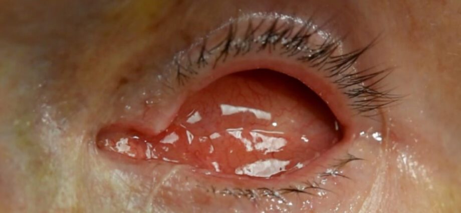
Sometimes it is necessary to remove the eye because it has an illness or has been badly damaged during trauma. This procedure is called enucleation. At the same time, it is associated with the placement of an implant, which will eventually accommodate an ocular prosthesis.
What is enucleation
Enucleation involves the surgical removal of the eye, or more exactly the eyeball. As a reminder, it is made up of different parts: the sclera, a hard envelope corresponding to the white of the eye, the cornea in the front, the lens, the iris, colored part of the eye, and in its center the pupil. Everything is protected by different tissues, the conjunctiva and Tenon’s capsule. The optic nerve allows the transmission of images to the brain. The eyeball is attached by small muscles within the orbit, a hollow part of the facial skeleton.
When the sclera is in good condition and there is no active intraocular lesion, the “table enucleation with evisceration” technique can be used. Only the eyeball is removed and replaced by a hydroxyapatite ball. The sclera, that is to say the white of the eye, is preserved.
How does an enucleation work?
The operation takes place under general anesthesia.
The eyeball is removed, and an intra-orbital implant is placed to accommodate the ocular prosthesis later. This implant is made either from a dermo-fatty graft taken during the operation, or from inert biomaterial. Where possible, the muscles for eye movement are attached to the implant, sometimes using a tissue graft to cover the implant. A shaper or jig (small plastic shell) is put in place while waiting for the future prosthesis, then the tissues covering the eye (Tenon’s capsule and conjunctiva) are sutured in front of the implant using absorbable stitches.
When to use enucleation?
Enucleation is offered in the event of an evolving lesion of the eye that cannot be treated otherwise, or when a traumatized eye endangers the healthy eye by sympathetic ophthalmia. This is the case in these different situations:
- trauma (car accident, accident in everyday life, fight, etc.) during which the eye may have been punctured or burned by a chemical product;
- severe glaucoma;
- retinoblastoma (retinal cancer mainly affecting children);
- ophthalmic melanoma;
- chronic inflammation of the eye that is resistant to treatment.
In the blind person, enucleation can be proposed when the eye is in the process of atrophy, causing pain and cosmetic modification.
After enucleation
Operative suites
They are marked by edema and pain lasting 3 to 4 days. An analgesic treatment makes it possible to limit the painful phenomena. Anti-inflammatory and / or antibiotic eye drops are usually prescribed for a few weeks. A week’s rest is recommended after the procedure.
The placement of the prosthesis
The prosthesis is placed after healing, ie 2 to 4 weeks after the operation. The installation, painless and not requiring surgery, can be performed in the ocularist’s office or in the hospital. The first prosthesis is temporary; the final one is asked a few months later.
Formerly in glass (the famous “glass eye”), this prosthesis is today in resin. Made by hand and made to measure, it is as close as possible to the natural eye, especially in terms of the color of the iris. Unfortunately, it does not allow to see.
The ocular prosthesis should be cleaned daily, polished twice a year and changed every 5 to 6 years.
Follow-up consultations are scheduled 1 week after the operation, then at 1, 3 and 6 months, then every year to ensure the absence of complications.
Complications
Complications are rare. Early complications include hemorrhage, hematoma, infection, scar disruption, implant expulsion. Others may occur later – conjunctival dehiscence (tear) in front of the implant, atrophy of the orbit fat with a hollow eye appearance, upper or lower eyelid drop, cysts – and require re-surgery.


