


Electrocardiograph: what is this medical instrument for?
The electrocardiograph records the electrical activity of the heart and assesses its state of health by detecting any abnormalities in its functioning. The examination performed, known as the electrocardiogram, is one of the essential heart examinations performed during any cardiology consultation.
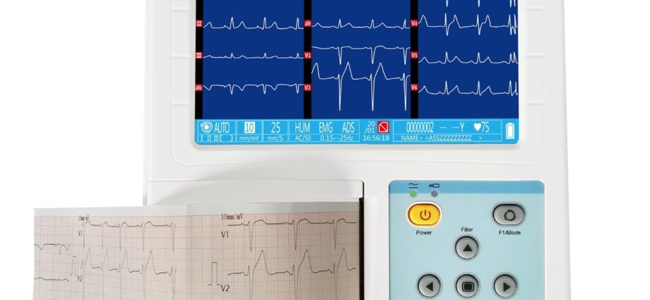
What is an EKG machine?
The activity of the heart is subjected to an electrical nerve impulse which induces its contraction and its relaxation in an automatic and periodic way. This nerve impulse, originating from the sinus node located at the top of the right atrium, is transmitted to neighboring heart muscle cells in the form of electrical waves which travel towards the tip of the heart (bottom left).
The electrocardiographs record these cardiac electrical waves and translate them into a curve, the analysis of which provides valuable information on the frequency and nature of the signals recorded and makes it possible to draw up a precise map of the heart and its working mechanics: this is the electrocardiogram (ECG).
Composition
Electrocardiographs are made up of 3 elements:
- the monitor, equipped with a screen, which records the cardiac electrical impulses;
- electrodes, disposable or reusable;
- cables to connect the electrodes to the monitor.
The different formats
Electrocardiographs exist in different formats:
- fixed in cabinet;
- portable on cart (7 to 10 kilograms);
- ultraportable (less than 1 kilogram and running on a rechargeable battery).
What is an EKG machine used for?
Deciphering the ECG allows the doctor to know the heart rate and to diagnose various pathologies linked to arrhythmias, a malformation of the heart, a physiological disorder or a heart disease:
- tachycardia;
- bradycardia;
- arrhythmia;
- extrasystole ;
- torsade de pointe;
- ventricular fibrillation ;
- ischemia;
- infarction;
- pericarditis (inflammation of the pericardium);
- valve disease (associated with atrial and / or ventricular hypertrophy);
- etc.
The ECG trace
The electrocardiograph records the heart’s electrical waves through electrodes placed on the patient’s skin at specific locations. The electrodes work in pairs. By varying the combinations of electrodes, we obtain different leads, 12 in all, which allow the ECG to be traced.
The ECG is a graph drawn on graph paper, the vertical axis of which corresponds to the amplitude of the electrical signal (with 1 mV = 1 cm) and the horizontal axis to its duration (1 sec = 25 mm). All charts are calibrated the same for comparison purposes.
Interpretation of the ECG
- The P wave is the first wave recorded: the electrical signal, coming from the sinus node, reaches the atria which contract to allow blood to pass to the ventricles;
- The following QRS complex is broken down into 3 waves: Q and S which symbolize the relaxation of the atria and their filling, and R which corresponds to the ventricular contraction allowing the ejection of blood towards the arteries. QRS also helps determine the electrical axis of the heart;
- The T wave is the last wave: it corresponds to the relaxation of the ventricles;
- The PQ segment is the time it takes for the electrical wave to travel from the atria to the ventricles: this is atrioventricular conduction;
- The ST segment represents the end of the ventricular contraction;
- The QT interval corresponds to the duration of the ventricular systole, that is to say a complete cycle of contraction / relaxation of the ventricles.
Heart rate is the number of QRS complexes per minute. It is normally 60 to 100 bpm (beats per minute) at rest.
ECG abnormalities
ECGs provide a wealth of information about the health of the heart. Changes in the duration, amplitude, direction of the waves and / or the appearance of additional signals are all signs of cardiac abnormalities.
In some cases, the cardiologist may also order an ambulatory Holter recording lasting 24 to 48 hours, during which the patient must note his periods of activity and rest, as well as any other information likely to shed light. interpretation of the ECG. The Holter may allow the detection of intermittent heart problems.
How is an EKG machine used?
The stages of operation
The examination, which is non-invasive and painless, lasts about 10 minutes. It can be performed in the hospital, at the office of the cardiologist or doctor, at home, or even outdoors by emergency physicians.
The patient is lying down with his arms at his sides, his legs extended. It should be relaxed to avoid electrical interference from the contraction of other muscles. The electrodes, coated with conductive gel, are positioned on the patient’s skin, which must be clean, dry and shaved if necessary to allow optimal adhesion. Their positioning obeys very precise rules:
- 4 frontal electrodes are placed at the wrists and ankles: they allow to know the electrical axis of the heart.
- 6 precordial electrodes are placed on the thorax: 2 to study the electrical activity of the right ventricle, 2 to study the interventricular wall and the tip of the heart, and 2 for the left ventricle.
Up to 18 electrodes can be placed to take an ECG. The placement points are always the same so that the produced ECGs can be compared.
When to use it?
The ECG can be done as a routine examination to check that the heart is functioning properly, as a follow-up examination during treatment, for a preoperative workup, or as a diagnostic examination when the patient complains of pain, dizziness or palpitations. cardiac.
An ECG can also be performed as part of a stress test, in an athlete for example. In this case, the patient must produce a sustained effort for 10 to 30 minutes. There are fewer electrodes and the respiratory rate and blood pressure are measured in parallel.
Precautions to take
There is no contraindication or specific patient preparation for performing an ECG.
The operator must ensure that the electrocardiograph is correctly adjusted: no interference, stable baseline, correct calibration (10 mm / mV), good paper flow speed (25 mm / sec), consistent trace ( electrodes must not be reversed).
How to choose an electrocardiograph?
The selection criteria
The use of electrocardiographs is restricted to medical personnel.
Several points should be considered when purchasing an electrocardiograph:
- sedentary or ambulatory use;
- use for measurements at rest or stress tests;
- screen: size, color, number of displayable tracks, touchscreen or not;
- printing of ECGs;
- power supply: mains, rechargeable battery, batteries;
- memory capacity for saving recordings;
- connectivity: Bluetooth connection, USB;
- existence of software dedicated to data interpretation;
- accessories: printing paper, sets of electrodes, cables, carrying case, etc. ;
- price: a few hundred to several thousand euros;
- verification of standards (CE marking).