


Contents
Definition of MRI in rheumatology
THEIRM is a diagnostic test that uses a magnetic field and radio waves to generate very precise 2D or 3D images of body parts or internal organs.
In rheumatology, a medical specialty which concernslocomotor device (diseases of the bones, joints and muscles), it finds a place of choice. It has even become essential in many rheumatological diagnoses, making it possible to obtain images that are much more precise than what is possible on an x-ray. MRI thus offers images of os, muscles, tendons, ligaments et cartilages.
Why perform an MRI in rheumatology?
The doctor may order an MRI to diagnose pathologies in the bones, muscles and joints. Thus the examination is carried out for:
- Understand the origin of persistent pain in the hips, shoulders, knees, ankles, back, etc.
- understand the intensification of pain during a Osteoarthritis
- evaluate the inflammatory rheumatism, and in particular the rheumatoid arthritis
- find the origin of pains and vascular disorders of the limbs.
The exam
The patient is placed on a narrow table capable of sliding into the cylindrical apparatus to which it is connected. The medical staff, placed in another room, manages the movements of the table on which the patient is placed using a remote control and communicates with him through a microphone.
Several series of cuts are made, according to all plans of the space. While the images are taken, the machine makes loud noises and the patient is asked not to move.
In some cases, a dye or contrast medium may be used. It is then injected into a vein before the exam.
What results can we expect from an MRI in rheumatology?
The images produced during the MRI will allow the doctor to make an accurate diagnosis of bone, muscle or joint diseases.
Thus, it will be able, for example, to detect:
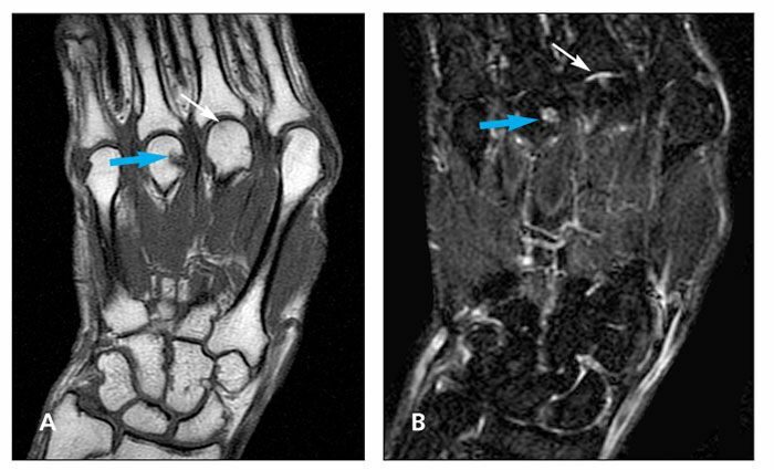
- in the case of arthritis : None synovites (inflammation of the synovium, membrane lining the inside of the capsule of mobile joints) and early erosions in locations that cannot be studied by ultrasound
- a cruciate ligament damage, Achilles tendon or knee cartilage
- a bone infection (osteomyelitis) or bone cancer
- a herniated disc, the spinal cord compression
- or algodystrophy or algoneurodystrophy: pain syndrome of a hand or a foot following a trauma such as a fracture