


Contents
Cochlea: all you need to know about this part of the ear
The cochlea is the part of the inner ear devoted to hearing. Thus, this spiral-shaped bone canal contains the organ of Corti, made up of hair cells that pick up different sound frequencies, from which these cells will produce a nerve message. Thanks to an auditory nerve fiber, the information will then be transmitted to the brain. In France, around 6,6% of the population has hearing loss, and this affects up to 65% of those over 70. This hearing loss can, in particular, be linked to exposure to too loud noises, which cause the destruction of hair cells in the cochlea, or even to advancing age, which reduces the number of hair cells in the ears. internal. Depending on the degree of hearing loss and the need for compensation, a cochlear implant may be offered, especially when hearing aids are not powerful enough to compensate for the deafness. In France, each year, 1 installations of this type are carried out.
Anatomy of the cochlea
Formerly called “snail”, the cochlea is the part of the inner ear that provides hearing. It is located in the temporal bone and owes its name to its spiral winding. Thus, the etymological origin of the term comes from the Latin “cochlea”, which means “snail”, and could, in imperial times, designate objects in the shape of a spiral. The cochlea is located in the last part of the inner ear where it is next to the labyrinth, the organ of balance.
The cochlea is made up of three canaliculi coiled in a spiral around a bony axis called the modiolus. It contains the organ of Corti, which is located between two of these canaliculi (that is, between the cochlear canal and the tympanic wall). This organ of Corti is a sensori-nervous organ, and one of the first anatomists to have described it was named Alfonso Corti (1822-1876). Made up of liquid as well as walls covered with internal and external hair cells located on its basilar membrane, the cochlea will transform the vibration of liquids and adjacent structures into a nervous message, and the information will be transmitted to the brain through the intermediary of a fiber of the auditory nerve.
Physiology of the cochlea
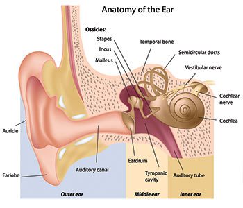
The cochlea plays a fundamental role in hearing, via the hair cells of the organ of Corti. In fact, the outer ear (which includes the auricular pinna whose role is to amplify the frequencies as well as the external auditory canal) ensures, with the middle ear, the conduction of sound towards the inner ear. And there, thanks to the cochlea, organ of this inner ear, the transmission of this message will be made to the cochlear neurons, which themselves will send it to the brain via the auditory nerve.
Thus, the principle of the functioning of hearing is as follows: when sounds are propagated in the air, this causes the clash of air molecules whose vibrations will be transmitted from the sound source to our eardrum, membrane located at the bottom of the external auditory canal. The tympanic membrane, vibrating like a drum, then transmits these vibrations to the three ossicles of the middle ear formed by the hammer, the anvil and the stirrup. Then, the vibration of the liquids induced by the caliper will then cause an activation of the hair cells, constituting the cochlea, thus creating bi-electric signals in the form of nerve impulses. These signals will then be transformed and decoded by our brain.
The hair cells, depending on their location in the cochlea, pick up different frequencies: in fact, those located at the entrance to the cochlea will resonate the high frequencies, while those located at the top of the cochlea, the bass frequencies.
Abnormalities, pathologies of the cochlea
The main anomalies and pathologies of the cochlea are linked to the fact that hair cells in humans do not regenerate once they have been damaged or destroyed. On the one hand, their exposure to too loud noises provokes their destruction. On the other hand, advancing age reduces the number of hair cells in the inner ears.
Acoustic overstimulation is therefore the cause of many physiological sequelae of the cochlea. These are induced by the activation of reactive oxygen species (or ROS, long considered toxic by-products of normal oxygen metabolism and involved in many abnormalities, but which researchers have recently shown that they were also involved in maintaining the balance of cells). These hearing deficits are also caused by apoptosis, the programmed death of hair cells.
More specifically, a scientific study carried out in 2016, in particular, demonstrated that the intracellular signaling of calcium (Ca2+) was involved in the initial pathophysiological mechanisms of the cochlea, following excessive exposure to noise. And so, it should be noted that the acoustic trauma generated by sound overstimulations occupies, today, the first rank of deafness factors.
The cochlear implant is a treatment indicated to establish an effective hearing in certain cases of bilateral profound deafness, and when conventional hearing aids are insufficient. The placement of such an implant must always be preceded by a prosthetic trial. The principle of this implant? Put in place in the cochlea a bundle of electrodes which will electrically stimulate the auditory nerve according to the frequency of the sounds which are picked up by the external part of the implant. In France, 1500 installations of this type are carried out each year.
Furthermore, the placement of a brainstem implant is also possible, in the case where the cochlear nerve is no longer functional, therefore preventing cochlear implantation. This deficiency of the cochlear nerve may be linked, in particular, to the removal of a local tumor or to an anatomical anomaly. These brainstem implants have, in fact, benefited from the technology developed for cochlear implants.
What diagnosis?
Deafness, also sometimes referred to as hearing loss, refers to decreased hearing acuity. There are rare cases of central deafness (involving the brain) but in the vast majority of cases, deafness is linked to a deficiency in the ear:
- conductive hearing loss is due to the outer or middle ear;
- Sensorineural hearing loss (also called sensorineural hearing loss) is caused by a failure in the inner ear.
Within these two categories, some deafness is genetic, while others are acquired.
A dysfunction of the inner ear, and therefore of the cochlea, is at the origin of sensorineural deafness (of perception): it generally reflects lesions of the hair cells or the auditory nerve.
The gold standard for assessing the level of noise audible to the ear is the audiogram. Carried out by an audiologist or hearing-aid acoustician, the audiogram will therefore allow the diagnosis of sensorineural hearing loss: this hearing test will assess the loss of hearing, but also quantify it.
History and anecdotes about the cochlea
It was in September 1976 that the first multi-electrode intracochlear implant was perfected, developed, patented and installed. It is, in fact, by continuing the French work of Djourno and Eyries that the doctor and surgeon specializing in otolaryngology Claude-Henri Chouard, assisted by his team from Saint-Antoine hospital, will invent this implant. Due to multiple economic but also industrial causes, the manufacture and marketing of cochlear implants have unfortunately, forty years later, completely escaped France. Thus, only four companies in the world now perform these tasks and they are Australian, Swiss, Austrian and Danish.
Finally, note: the cochlea, among all its virtues, has one less known, but very useful to archaeologists: it can indeed help them determine the sex of a skeleton. The cochlea is located in the hardest bone of the skull – the rock of the temporal bone -, and it will be possible, by means of a specific archaeological technique, to establish, thanks to it, the sex of very ancient, whether fossil or not. And this, even when it comes to fragments.