


Contents
Coccyx
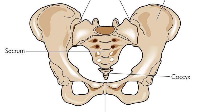
The tailbone (from the Greek kokkuks), located under the sacrum, is the bone of the final part of the spine. It helps to carry the weight of the body.
Anatomy of the tailbone
The tailbone a bone in the lower part of the spine. It constitutes its extremity but does not harbor bone marrow. It has a triangular shape, the point of which is directed downwards and is found at the level of the anus. Located under the sacrum, it also forms with the latter the posterior part of the bony pelvis.
It is made up of three to five small, irregular coccygeal vertebrae joined together by joints and ligaments. It is a remnant of the mammalian tail.
Physiology of the coccyx
The tailbone supports the spine and thus contributes to the axial support of the body.
Associated with the hip bones and the sacrum, the coccyx also constitutes the pelvis which has the main role of supporting the weight of the upper body.
Pathologies of the coccyx
Coccyx fracture : most often occurs following a heavy fall on the buttocks, but can also be caused by childbirth (mechanical crushing due to the passage of the baby), a disease which weakens the bones (osteoporosis) or even mechanical stress imposed on the baby. coccyx. This fracture causes in all cases a sharp pain which interferes with the sitting position. Usually rest and taking painkillers and anti-inflammatory drugs are sufficient for healing. Very painful fracture, it is recommended to sit on a suitable cushion such as a buoy or a hollow cushion. In some very rare cases, the fracture is accompanied by a deviation of the bone. It must then be replaced by an intervention under general anesthesia.
Coccygodynie : persistent pain in the tailbone, exacerbated when sitting or standing (5). The causes, often traumatic, can be multiple: a fracture, a fall with a severe shock, a bad or prolonged sitting position (e.g. driving), childbirth, a disease (osteoporosis), a coccygeal spine, a dislocation, arthritis… A study (6) also shows a link between coccygodynia and depression. If the pain is not treated, it can quickly become disabling for people who suffer from it (sitting or even standing too painful).
Epine coccygienne : bone growth present at the tip of the coccyx which represents 15% of cases of coccygodynia. The spine exerts pressure in a seated position and causes pain and inflammation of the tissues under the skin.
Luxation coccygienne : dislocation which concerns the joint between the sacrum and the coccyx or the discs of the coccyx itself. It is very common (20 to 25% of cases of tailbone pain).
Calcification : it is possible that a small calcification appears in a disc between the vertebrae. This presence results in a sudden and very intense pain making it impossible to sit down. An anti-inflammatory treatment for a few days is effective.
Pilonidal cyst : subcutaneous cyst which forms in the inter-gluteal fold, at the level of the end of the coccyx. It is a hair that grows under the skin which eventually becomes infected: it is the abscess, a pocket of pus forms. In these cases, surgery is necessary. A congenital pathology, it affects men up to 75% (7). It could also be caused by the friction of the hairs of the inter-gluteal fold which would be enough to pierce the skin and form a cyst. This could explain the frequency of cysts in people with heavy hairiness or overweight.
Recurrences are not uncommon because the pocket formed by the cyst still exists after the operation.
Treatments and prevention of the coccyx
The elderly represent a population at risk of coccyx fractures because they are more exposed to falls and their bones are more friable. The same is true for people with osteoporosis. Preventing a fall is not easy, but it is advisable to consume foods rich in calcium and vitamin D in order to strengthen the bones and reduce the risk of fractures.
Health professionals advise adopting a good way of sitting: choose a comfortable seat when possible and avoid sitting for a long time. Long trips by car are not recommended, but if they do, a buoy or hollowed-out cushion can prevent the pain. For athletes, cycling and horse riding is not recommended.
Tailbone exams
Clinical examination: carried out by the doctor, it first includes the questioning (general, on the causes of the accident or a history). It is followed by a physical examination of the coccyx (inspection and palpation) which will be completed by an examination of the lumbar, pelvis and lower limbs.
Radiography: a medical imaging technique that uses x-rays. Radiography is the gold standard examination indicated in all patients with tailbone pain. A standing, lateral x-ray mainly detects fractures.
Bone scintigraphy: imaging technique which consists of administering to the patient a radioactive tracer which spreads in the body or in the organs to be examined. Thus, it is the patient who “emits” the radiation that will be picked up by the device. The scintigraphy makes it possible to observe the bones and the joints. In cases of the coccyx, it is mainly used in conjunction with radiography for the diagnosis of stress fractures.
MRI (magnetic resonance imaging): medical examination for diagnostic purposes carried out using a large cylindrical device in which a magnetic field and radio waves are produced. It can highlight an inflammation of the coccyx region or the consequences of a dislocation or can rule out certain pathologies, for example.
Infiltration: it can be performed as part of a treatment for tailbone pain. It consists of injecting between the discs of the vertebrae local anesthetics and corticosteroids. The results are satisfactory in 70% of cases (2).
Coccygectomy: Surgery that removes segments of the tailbone. It may be offered to some people with chronic coccygodynia who are refractory to treatment. The results are good and excellent in 90% of cases (3) but there are risks of complications such as wound infection. The improvement is felt after two or three months, or even more.
Anecdote and coccyx
The tailbone owes its name to the Egyptian cuckoo clock, the Clamator Glandarius, due to its resemblance to the bird’s beak. It was Herophilus, a Greek doctor who lived in Alexandria, who named him so. Cuckoo saying kokkyx in Greek.