


Contents
3D ultrasound in practice
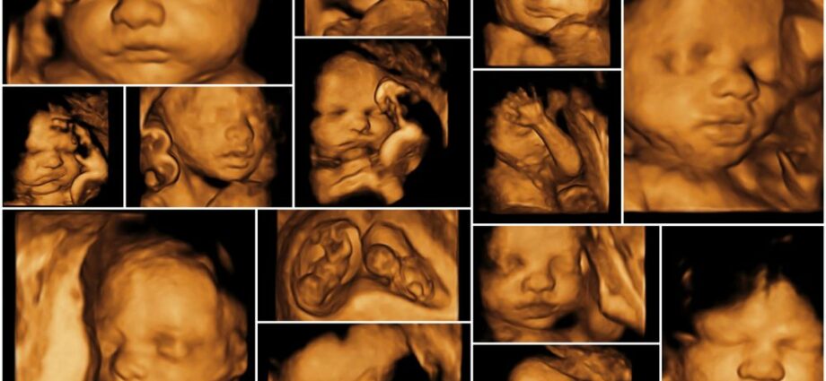
Ultrasound is always a moment of emotion, all the more so when the doctor suggests that we discover our baby’s face “for real”. He then reproduces before our tender eyes images of our child in 3D. Magnificent pictures will proclaim some, strange will say others. In any case, the emotional impact of these images is immense and often takes precedence over the purely medical aspect.
3D ultrasound: what is changing?
Three ultrasounds are recommended in the classic follow-up of pregnancy. They take place in the first, second and third trimester respectively and are carried out in 2D. As a reminder, ultrasound is a medical imaging technique that uses high frequency ultrasonic waves to produce images of the baby inside the uterus. The goal of fetal ultrasounds is to monitor your baby’s growth and look for any deformities. 3D ultrasound has grown significantly in recent years. It uses the same operating principle and has the same functionalities as 2D. The device makes it possible to reconstruct volume images of the explored area from the ultrasonic waves reflected in the mother’s body. Some ultrasounds have the ability to provide real-time 3D images. These are the 4D ultrasounds.
3D ultrasound indications
This technique undoubtedly improves the ultrasound rendering by giving a three-dimensional image, (more or less) faithful reproduction of the baby. Many professionals agree to show these beautiful images to parents who wish at the end of the exam, as a souvenir, but without medical purpose. 3D reconstruction makes it possible to see certain organs with more precision. However, says Dr Sahar Kaddioui Maalej, obstetrician-gynecologist and sonographer, “ she has a very limited medical interest in pregnancy monitoring »And is indicated in very specific cases. ” Most abnormalities are detected by 2D ultrasound. 3D is in a way an additional step in the early diagnosis of certain malformations in the fetus », She specifies. Indeed, this technology is a means of diagnosing some pathologies of the skeleton (cleft lip), the skull or the face (facial dysmorphia). The 3D volume reconstruction also allows you to see precisely the vessels, heart and reproductive system of the baby. As with the 2D ultrasound, the specialist spreads a watery gel on the skin and sweeps the stomach with a probe. To get quality pictures, the baby must be in the right position. What is not always obvious, the fetus sometimes hides its face with its hands, sticks to the placenta or shows its back to the lens.
In video: The clear egg is rare, but it does exist
Watch out for commercial ultrasounds!
More and more private practices are offering 3 D ultrasounds, as a “souvenir”. Parents who wish can leave with photos and even a film of their baby, after paying a large sum. The French College of Gynecologists and Obstetricians has repeatedly warned couples who would be tempted by this experience. These ultrasounds can lead to too long exposure of the fetus to ultrasound.. And they are not always performed by professionals who have been trained in fetal ultrasound. ” We are no longer in medicine, insists Dr Kaddioui Maalej. These ultrasounds have no medical purpose, unlike the three other examinations recommended in pregnancy monitoring..