


Contents
Sternum
The sternum (from Latin sternum, from Greek sternon) is a bone of the thorax which constitutes the rib cage on its middle portion.
Anatomy of the breastbone
The sternum is a flat bone located in front of the thorax, in the midline of the body (in the middle). It articulates on each side with the first seven ribs as well as with the clavicles at which it forms the sternoclavicular joint. Placed on the surface under the skin, it is located in front of a large part of the heart.
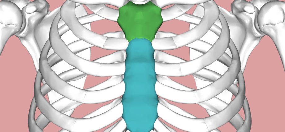
The breastbone is made from the fusion of three bony pieces:
- Le handle sternal,
- The body of the breastbone,
- The xiphoid process.
There are three important anatomical landmarks:
- The jugular notch marks the upper edge of the sternum. It is easily palpable under the skin, it is the hollow that we feel at the base of the neck.
- The sternal angle is at the border of the sternal manubrium and the body. Also palpable, it stands out in the form of a horizontal ridge.
- The lower sternal joint, which is located at the junction between the body of the sternum and the xiphoid process.
Physiology of the breastbone
The sternum participates in the formation of the bone structure of the rib cage. The ribs and thoracic vertebrae combine with it to complete it.
Pathologies of the sternum
Sternum fracture :
Sternum fractures are associated with trauma, whether direct or indirect. A direct impact can be due to a car accident (seat belt pressing on the chest or impact of a steering wheel) or related to sports. Indirect causes of fractures can occur spontaneously in elderly people with osteoporosis, for example. Stress fractures have also been identified in athletes following repetitive upper body exercises. These breastbone fractures can either happen in isolation or be associated with other injuries:
– Isolated: only the sternum is affected. The majority of patients recover completely after several weeks of convalescence.
– Associated with other injuries: two thirds of sternum fractures are associated with serious pathologies that can cause death in 25 to 45% of cases (3). These injuries can only affect the tissues or reach deeper into the rib cage (rib fractures, heart, lung and spine damage, etc.).
Sternoclavicular dislocation : dislocation of the joint between the clavicle and the sternum, it is four times less frequent than the acromioclavicular.
Chest pain : they have multiple causes and can sometimes be felt in the sternum. These pains are generally due to heart disease (eg myocardial infarction) or vascular disease (eg pulmonary embolism) and require rapid medical treatment.
Sternal fente : rare malformation of the sternum, of unknown cause. During embryonic life, it results in a defect in the fusion of the bone bars intended to constitute the sternum, which normally takes place from top to bottom to close it completely. Surgery during the first weeks after birth closes the breastbone and thus protects the heart and the large vessels behind it.
Sternocostoclavicular hyperostosis : rare pathology of unknown cause, it results in hypertrophy and condensation of the sternum, collarbones and first ribs. It preferentially affects the middle-aged man. The main symptom is painful swelling in the breastbone.
Tumors of the breastbone : Bone tumors of the chest wall can very rarely be located on the breastbone or collarbone. This type of bone tumor represents less than 5% of all bone tumors (6).
Prevention of breastbone
Pathologies of the sternum are due to external trauma or rare diseases of unknown causes. It therefore seems difficult to prevent them.
Sternum examinations
Sternal puncture: practice of inserting a needle into the breastbone to remove bone marrow. This marrow contains the so-called hematopoietic cells, which are at the origin of the various blood cells. The laboratory analysis of these cells is the myelogram. It is used to diagnose an abnormality in one of the blood cell lines. This puncture can also be carried out in the bone of the pelvis, it is then a lumbar puncture.
Imaging exams:
- Radiography: a medical imaging technique that uses X-rays. Radiography of the sternum or sternoclavicular joints is a standard examination of reference in pathologies linked to trauma.
- Scanner: imaging technique which consists of “scanning” a given region of the body in order to create cross-sectional images, thanks to the use of an X-ray beam. We also speak of computed tomography or CT scans. This exam allows a good visualization of the medullary bone as well as the soft tissues of the joint and around the joint.
- MRI (magnetic resonance imaging): medical examination for diagnostic purposes carried out using a large cylindrical device in which a magnetic field and radio waves are produced. It provides very precise images of the mineralized bone of the sternum.
- Bone scintigraphy: imaging technique which consists of administering to the patient a radioactive tracer which spreads in the body or in the organs to be examined. Thus, it is the patient who “emits” the radiation that will be picked up by the device. The scintigraphy makes it possible to observe the bones and the joints. In cases of the sternum, it is used in particular for the diagnosis of sternocosto-clavicular hyperostosis.
History and symbolism of the sternum
It is estimated that 5% of the world’s population has a “sternal form”, or sternal perforation, or a round opening on the body of the breastbone. This hole, similar to the one left by a bullet passing through the breastbone, is actually explained by a defect in ossification (8,9).