


Contents
Rib cage
The rib cage (from the Greek thôrax, chest) is an osteo-cartilaginous structure, located at the level of the thorax, which participates in particular in the protection of vital organs.
Thoracic anatomy
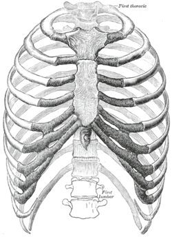
Structure of the rib cage. It is made up of different elements (1) (2):
- The breastbone which is a long, flat bone located in the front and center.
- The thoracic spine, located at the back, which is made up of twelve vertebrae, themselves separated by the intervertebral discs.
- The ribs, twenty-four in number, which are long and curved bones, going from the back to the front through the lateral face.
Shape of the rib cage. The ribs start from the spine and are attached to the breastbone by the costal cartilage, with the exception of the last two lower ribs. Called floating ribs, these are not attached to the sternum (1) (2). These junctions make it possible to give the structure in the form of a cage.
Intercostal spaces. Eleven intercostal spaces separate the twelve ribs on a lateral face. These spaces are made up of muscles, arteries, veins, as well as nerves (2).
Thoracic cavity. It contains various vital organs including the heart and lungs (2). The base of the cavity is closed by the diaphragm.
Functions of the rib cage
Protective role of internal organs. Due to its shape and constitution, the rib cage protects vital organs like the heart and lungs, as well as some abdominal organs (2).
Mobility role. Its partly cartilaginous constitution gives it a flexible structure allowing it to follow the movements of the spine (2).
Role in respiration. The flexible structure of the cage, as well as the different joints gives it large amplitudes of movement, participating in the respiratory mechanics. Various breathing muscles are also located in the rib cage (2).
Pathologies of the rib cage
Thoracic trauma. It corresponds to the damage to the thoracic cage due to a shock to the thorax (3).
- Fractures. The ribs, the sternum as well as the dorsal spine can undergo various fractures.
- Thoracic flap. It corresponds to a segment of the chest wall that has separated and follows fractures of several ribs (4). This leads to respiratory complications with paradoxical breathing.
Deformities of the chest wall. Among these deformations, we find that of the anterior thoracic wall:
- The thorax in a funnel, causing a hollow deformation, due to a projection behind the sternum (5).
- The thorax keeled, causing a deformity in bump, due to a projection forward of the sternum (5) (6).
Pneumothorax. It refers to the pathology affecting the pleural cavity, the space between the lungs and the rib cage. It is manifested by severe chest pain, sometimes associated with difficulty breathing.
Tumors of the chest wall. Primary or secondary tumors can develop in bone or soft tissue (7) (8).
Maladies of the os. The rib cage can be the site of the development of bone diseases such as osteoporosis or ankylosing spondylitis.
Rib cage treatments
Medical treatment. Depending on the trauma or pathology, analgesics and anti-inflammatory drugs may be prescribed.
Surgical treatment. Surgery may be performed for chest wall deformities, chest trauma, as well as for tumors (5) (7) (8).
Thoracic cage exams
Physical examination. Diagnosis begins with a physical examination to assess the symptoms and characteristics of the pain.
Medical imaging exams. Depending on the suspected or proven pathology, additional examinations may be performed such as an x-ray, an ultrasound, a CT scan, an MRI or a scintigraphy (3).
History and symbolism of the rib cage
Chest compression, used today as a first aid procedure, was first described in animals in 18749 before being demonstrated in humans in 1960 (10).