Contents

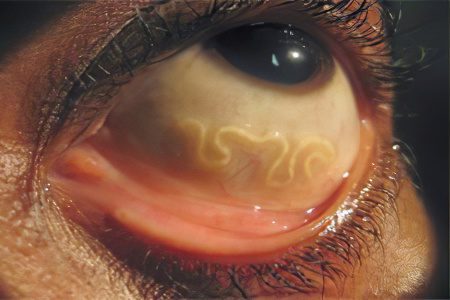
Ocular toxocariasis is a lesion of the organ of vision by migrating larvae of the helminth worms Toxocara canis. The natural host of toxocara are representatives of the canine family (dogs, wolves, jackals), less often cats are carriers of worms. The larvae do not seek to enter the human body, and infection occurs most often by accident. It is not able to develop into an adult toxocara in the human body.
Eye damage by toxocara larvae is observed quite rarely, no more than 9% of all cases of toxocariasis recorded in humans.
Ocular toxocariasis develops most often when a small number of larvae enter the human body. No more than one larva is predominantly found in the eye. The vast majority of cases are older people, although children are also susceptible to invasion.
All cases of ocular toxocariasis are usually divided into 2 basic groups:
Chronic endophthalmitis with exudation;
Solitary granulomas.
The first cases of ocular toxocariasis were described not so long ago, in the 50s of the last century. Moreover, patients were admitted to ophthalmologists with a diagnosis of retinoblastoma and Coates’ disease. However, later they found nematode larvae and hyaline capsules.
Causes of ocular toxocariasis

The causes of ocular toxocariasis lie in the entry of a worm larva into the human body and in its further migration to the organ of vision. Infection with toxocara occurs by the fecal-oral route.
Possible sources of invasion:
Soil containing helminth eggs. It can be introduced into the human gastrointestinal tract if the rules of personal hygiene are not followed, by eating poorly processed berries, vegetables, fruits.
With some psychological diseases, for example, when people eat the earth for food.
Toxocara eggs are carried by dogs, including puppies. In addition to being found in their feces, the eggs are often attached to their fur. Therefore, contact with animals can be dangerous, especially if hands are not thoroughly washed with soap and water after contact.
Toxocara eggs are carried by cockroaches. They eat them, after which they seed human food with viable eggs.
In the risk group for infection with ocular toxocariasis are veterinarians, employees of reception centers for animals, children aged 3 to 5 years, hunters, summer residents, gardeners and other farmers.
In the body of a dog, toxocara migrates according to the scheme: gastrointestinal tract> portal vein> liver> right atrium> lungs> trachea> larynx> esophagus> stomach> intestines, where toxocara eventually turns into a sexually mature individual. In the human body, there are no conditions for the worm to complete its life cycle. Therefore, toxocara migrates in the human body until it stops in one or another organ under the influence of the immune system, which will not allow it to move further. After that, the larva of the worm creates a dense capsule around itself and becomes inactive for a long time. However, in the organ where it stopped, chronic inflammation begins. In this case, we are talking about the eyes.
Symptoms of ocular toxocariasis

Symptoms of ocular toxocariasis in humans may include the following:
A sharp deterioration in vision, up to its partial or complete loss. In most cases, only one eye is affected.
Protrusion of the eyeball from the orbit.
Severe hyperemia of the conjunctiva.
Redness of the eyelids, their swelling.
Severe pain in the eyes of a bursting character.
Strabismus.
Blurred vision.
When the organ of vision is damaged by worm larvae, a person has granulomas in the posterior part of the eye, chronic uevitis, endophthalmitis, keratitis, optic neuritis, migrating larvae in the vitreous body.
In childhood, ocular toxocariasis manifests itself mainly in strabismus; when the larva moves, the patient develops a migrating scotoma.
Treatment of ocular toxocariasis

Treatment of ocular toxocariasis in both children and adults is reduced to the appointment of subconjunctival injections of the steroid drug Depo-Medrol. This glucocorticosteroid drug allows you to remove inflammation from the eyeball and eliminate the worm larva. However, the mainstay of treatment is usually surgery. With retinal detachment, laser correction is indicated.
For the first time, a 13-year-old girl was successfully treated for ocular toxocariasis back in 1968. She received subconjunctival injections of Depo-Medrol. However, in some cases, the effect of this drug is absent. Therefore, doctors recommend implementing the same scheme for the treatment of ocular toxocariasis as for getting rid of visceral parasitic invasion. That is, patients are prescribed a standard anthelmintic regimen consisting of combination therapy.
To rid the patient of toxocariasis, Mintezol (Thiabendazole), Vermox (Mebendazole) and Ditrazine (Diethylcarbamazine) are prescribed. The dose is calculated depending on the body weight of the person.
There is also information about the successful deliverance of patients from ocular toxocariasis using photo- and laser coagulation. These procedures allow destroying toxocary granulomas (the membranes in which the larvae of the worms are located).
As for the prognosis for recovery, it depends on the severity of the disturbances caused by the parasite larva and on the duration of the invasion.


