


Contents
Ocular hypertension: interpretation and treatment
There are few or no symptoms of chronically elevated eye pressure. Nausea, pain or vomiting can sometimes occur in case of sudden or very sharp rise in eye pressure. However, the pathology should be taken seriously because of its consequences on vision.
What is ocular hypertension
It is a pathology that affects about 4 to 7% of the population and its frequency increases with age.
“Ocular hypertension is characterized by an increase in the pressure exerted by the aqueous humor, which is the liquid located between the cornea and the lens,” explains Dr. Cédric Lamirel, ophthalmologist at the Rothschild Foundation hospital. This liquid is renewed every two to three hours. This ocular hypertension comes from a poor evacuation of the aqueous humor.
Who is concerned ?
Common after 40 years, ocular hypertension is the first risk factor for glaucoma. Taken in time, it can fortunately be effectively controlled provided that regular checks are carried out.
How is ocular hypertension measured?
Intraocular pressure is measured in millimeters of mercury (“mmHg”) by an ophthalmologist. The pressure measurement is always done through the cornea and must be interpreted according to the central thickness of the cornea.
Eye pressure measurement
“In most cases, the device used to measure this pressure is the forced air tonometer,” continues the specialist. It projects a jet of air on the cornea creating a temporary deformation. The value of the intraocular pressure is obtained by measuring the pressure of the air jet required to flatten the cornea. There is no contact with the cornea and therefore no anesthesia necessary ”.
Using the applanation tonometer
Another option: the applanation tonometer which requires the use of anesthetic eye drops beforehand. “Once anesthetized, the measuring device comes into direct contact with the cornea to temporarily flatten the cornea and measure intraocular pressure. This is the reference method ”.
Bi-digital palpation of the eye
Finally, the bi-digital palpation of the eye through the closed eyelids makes it possible to roughly estimate whether there is a strong ocular hypertonia or not: it is only used in patients who cannot be installed in front of the devices. of ophthalmologist measurements.
Interpretation of eye pressure
“Eye pressure is normal between 10 and 21 millimeters of mercury, but this figure can vary with the age of the subject, the position of the patient, the thickness of the cornea or even according to the hours of the day or night. It should be roughly equal in both eyes, ”adds the opthalmologist. Beyond 21 mmHg, there is ocular hypertonia.
Symptoms and causes of high eye pressure
Symptoms of eye pressure
Nausea, pain or vomiting can sometimes occur in the event of a sudden and very sharp rise in eye pressure.
Most of the time, there are few or no symptoms of chronically high eye pressure. However, the pathology should be taken seriously because of its consequences on vision.
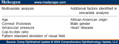
“When the pressure is too high, it can lead to deterioration of the fibers of the optic nerve, which constitutes glaucoma,” recalls our interlocutor. This eye disease causes a progressive amputation of the visual field which can lead to blindness if it is not detected in time. The risk of developing glaucoma depends on the extent of this hypertonia.
“But also other factors such as the thickness of the cornea, the shape of the optic nerve, the visual field, the presence of high myopia, a family history of glaucoma, etc. Estimating this risk makes it possible to offer the patient either regular monitoring if the risk is low, or treatment to prevent the onset of glaucoma if the risk is high ”.
Causes of eye strain
Some of the main causes that lead to ocular hypertension include:
- Insufficient drainage of aqueous humor;
- The effects of certain drugs, especially those that contain corticosteroids;
- Trauma to the eye;
- Eye diseases such as pseudo-exfoliative syndrome or pigment dispersion syndrome which tend to increase tension in the eyes.
Eye pressure treatments
Administration of eye drops
Treatment will consist of the daily administration of eye drops, usually for life. These ocular hypotensive drugs are intended to decrease the production of aqueous humor or to increase its drainage.
“If the medical treatment is insufficient, surgery and / or laser treatment can be offered by the ophthalmologist,” says Dr. Lamirel.
Surgical procedures
The trabéculoplastie
It is performed by a laser beam applied to the trabeculum, which increases its capacity to evacuate aqueous humor.
Non-perforating deep sclerectomy and trabeculectomy
These are two surgical techniques that allow the aqueous humor to be evacuated under the conjunctiva (the filtration bubble)
Iridoplasty and iridotomy
These are laser treatments of the iris to facilitate the flow of aqueous humor between different parts of the eye.