


Contents
Morphological ultrasound: the 2nd ultrasound
The second pregnancy ultrasound, called morphological ultrasound, is an important step in pregnancy monitoring because it can detect possible fetal malformations. For parents, it is also a highlight: that of discovering the sex of the baby.
The second ultrasound: when does it take place?
The second ultrasound takes place on the 5th of pregnancy, between 21 and 24 weeks old, ideally at 22 weeks old.
It is not compulsory but is part of the examinations systematically prescribed during pregnancy follow-up and highly recommended.
The course of the ultrasound
For this test, it is not necessary to be fasting or to have a full bladder. On the other hand, it is not recommended to put cream or oil on the stomach during the 48 hours preceding the ultrasound so as not to affect the quality of the image.
The practitioner coats the belly of the mother-to-be with gelled water to facilitate the passage of ultrasound. Then, he will move the probe on the stomach in order to obtain different images, or sections, of the baby. This second ultrasound lasts a little longer than the first because it methodically studies the full anatomy of the baby.
Why is it called morphological ultrasound?
The main objective of this ultrasound is to look for morphological abnormalities. The practitioner will methodically study each organ by making transverse sections which allow, at each “level”, to control the presence and shape of the different organs: the heart, the brain, the different organs of the abdomen (stomach, bladder, intestine), all four limbs.
It is during this examination that fetal malformations are most easily detected. However, although it is more and more efficient and sophisticated, morphological ultrasound is not 100% reliable. It sometimes happens that a fetal anomaly, even present at this stage of pregnancy, is not detected during this ultrasound. This happens when the malformation is not or hardly accessible in the image, the position of the fetus masks the malformation, or when the future mother is overweight. Subcutaneous adipose tissue can in fact interfere with the passage of ultrasound and alter the quality of the image.
During this second ultrasound, the practitioner also checks:
- baby growth using biometrics (measurement of biparietal diameter, cranial perimeter, abdominal perimeter, femoral length, transverse abdominal diameter) the results of which will be compared to a growth curve;
- the placenta (thickness, structure, level of insertion);
- the amount of amniotic fluid;
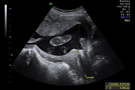
- the internal opening of the cervix in particular in the event of contractions.
It is also during this second ultrasound that the announcement of the baby’s sex takes place – if the parents wish to know it of course – and if the baby is well positioned. At this stage of pregnancy, the external genitalia are formed and recognizable in the image, but there is always a small margin of error, depending on the position of the baby in particular.
A Doppler is sometimes performed during this ultrasound. With sounds transcribed on a graph, it helps control blood flow in different vessels and arteries (uterine arteries, umbilical arteries, cerebral arteries). It is a complementary tool for controlling fetal growth in certain risky situations or obstetric complications (1):
- gestational diabetes;
- hypertension;
- fetal distress;
- growth retardation in utero (IUGR);
- an abnormality of the amniotic fluid (oligoamnios, hydramnios);
- fetal malformation;
- a monochorial pregnancy (twin pregnancy with a single placenta);
- pre-existing maternal disease (hypertension, lupus, nephropathy);
- a history of obstetric vascular pathologies (IUGR, pre-eclampsia, placental abruption);
- a history of death in utero.
The fetus at the time of the 2nd ultrasound
At this stage of pregnancy, the baby is about 25 cm from head to toe, half of his birth size. It weighs only 500 gr. Its feet are approximately 4 cm (2).
He still has a lot of room to move, even if the mother-to-be does not always feel these movements. He cannot see but he is very sensitive to touch. He sleeps about 20 hours a day.
Her legs, her arms show up clearly, and even her hands with well-formed fingers. In profile, the shape of his nose emerges. Its heart is the size of an olive, and within it all four parts are present as are the pulmonary artery and aorta.
We see almost all the vertebrae which in the image, form a kind of stop. He has no hair yet, but a simple down.
For parents, this second ultrasound is often the most pleasant: the baby is large enough so that we can clearly see his face, his hands, his legs, but still small enough to appear in full on the screen and allow a overview of this little being already well formed.
The problems that the 2nd ultrasound can reveal
When a morphological abnormality is suspected, the mother-to-be is referred to an antenatal diagnosis center and / or a reference sonographer. Other examinations are performed to confirm the anomaly and refine the diagnosis: amniocentesis, MRI, cardiac ultrasound, MRI or fetal scan, fetal blood puncture, blood tests for the couple, etc.
Sometimes examinations do not confirm the anomaly. Pregnancy monitoring then resumes normally.
When the anomaly detected is less serious, a specific follow-up will be set up for the remainder of the pregnancy. If the anomaly can be treated, particularly surgically, from birth or during the first months of life, everything will be organized to implement this care.
When the prenatal diagnosis confirms that the baby is suffering from “a condition of particular gravity recognized as incurable at the time of diagnosis” according to the texts, the law (3) authorizes patients to request a medical termination of pregnancy (IMG) or “therapeutic abortion” at any term of pregnancy. Specific structures approved by the Biomedicine Agency, the Multidisciplinary Centers for Prenatal Diagnosis (CPDPN), are responsible for certifying the severity and incurability of certain fetal pathologies and thus authorizing IMG. These are genetic diseases, chromosomal abnormalities, malformation syndromes or very serious anomaly (of the brain, heart, absence of kidneys) inoperable at birth and which can lead to the death of the baby at birth or in his early years. , infection which could prevent the survival of the baby or cause his death at birth or in his first years, pathology leading to severe physical or intellectual disability.
During this second ultrasound, other pregnancy complications can be detected:
- intrauterine growth retardation (IUGR). Regular growth monitoring and a Doppler ultrasound will then be performed;
- a placental insertion abnormality, such as a placenta praevia. An ultrasound will monitor the evolution of the placenta.