


Contents
lungs
The lungs (from Latin pulmo, -onis) are structures of the respiratory system, located within the rib cage.
Lung anatomy
Position. Two in number, the lungs are located in the thorax, more particularly within the thoracic cage where they occupy most of it. The two lungs, right and left, are separated by the mediastinum, located in the center and composed in particular of the heart (1) (2).
Pleural cavity. Each lung is surrounded by the pleural cavity (3), which is formed from two membranes:
- an internal layer, in contact with the lung, called pulmonary pleura;
- an external layer, in contact with the chest wall, called the parietal pleura.
This cavity is composed of a serous fluid, the transudate, allowing the lung to slide. The set also helps to maintain the lung and prevent it from sagging.
Overall structure of the lungs. The right and left lungs are connected by the bronchi and the trachea.
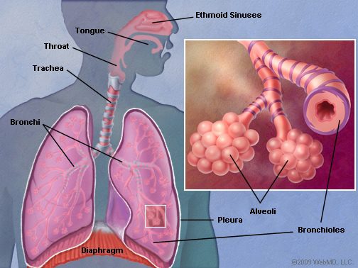
- Trachea. The trachea, respiratory duct coming from the larynx, passes between the two lungs on their upper parts and separates into two right and left bronchi.
- Bronchi. Each bronchus is inserted at the level of a lung. Within the lung, the bronchi divide to form smaller and smaller structures up to the terminal bronchioles.
Pyramidal in shape, the lungs have several faces:
- An external face, contiguous to the costal grill;
- An internal face, where the bronchi are inserted and the blood vessels circulate;
- A base, resting on the diaphragm.
The lungs are also made up of lobes, separated by fissures: two for the left lung and three for the right lung (2).
Lobe structure. Each lobe is made up and functions like a small lung. They contain branches of bronchi as well as pulmonary arteries and veins. The endings of the bronchi, called terminal bronchioles, form a sac: the acinus. The latter is made up of several dents: the pulmonary alveoli. The acinus has a very thin wall in contact with the air coming from the bronchioles and the network formed by the pulmonary capillary vessels (2).
Double vascularization. The lungs receive a double vascularization:
- a functional vascularization constituted by the network of pulmonary arteries and veins, making it possible to oxygenate the blood;
- a nutritive vascularization constituted by the bronchial arteries and veins, making it possible to provide the essential elements for the proper functioning of the lungs (2).
Respiratory system
The lungs play an essential role in breathing and oxygenating the blood.
Pulmonary pathologies and diseases
Pneumothorax. This pathology corresponds to an abnormal entry of air into the pleural cavity, the space between the lungs and the rib cage. It manifests as severe chest pain, sometimes associated with difficulty breathing (3).
Pneumonia. This condition is an acute respiratory infection directly affecting the lungs. The alveoli are affected and become filled with pus and fluid, causing breathing problems. Infection can especially be caused by bacteria, viruses or fungi (4).
TB. This disease corresponds to a bacterial infection often found in the lungs. Symptoms are chronic cough with bloodshed, intense fevers with night sweats, and weight loss (5).
Acute bronchitis. This pathology is due to an infection, often viral, in the bronchi. Frequent in winter, it causes coughs and fever.
Lung cancer. Malignant tumor cells can develop in the lungs and bronchi. This type of cancer is one of the most common in the world (6).
Treatments
Medical treatment. Depending on the pathology diagnosed, different treatments may be prescribed such as antibiotics or analgesics.
Surgical treatment. Depending on the pathology diagnosed, surgery may be necessary.
Exploration and exams
Physical examination. An analysis of the breath, breath, lungs and symptoms perceived by the patient is performed in order to assess the pathology.
Medical imaging exam. Lung radiology, chest CT, MRI or lung scintigraphy can be done to confirm a diagnosis.
Medical analysis. In order to identify certain pathologies, blood tests or analyzes of pulmonary secretions, such as cytobacteriological examination of sputum (ECBC), may be performed.
History
Discovery of tuberculosis. Tuberculosis is a pathology known since Antiquity and was notably described by Hippocrates. However, the pathogen responsible for this disease was not identified until 1882 by the German physician Robert Koch. He described a bacterium, and more particularly a tubercle bacillus, called Koch’s bacillus or Mycobacterium tuberculosis (5).