


Contents
Leg
The leg (from the Latin gamba meaning hock of animals) is a part of the lower limb located between the knee and the ankle.
Anatomy of the legs
Leg skeleton. The leg is made up of two bones linked together by a bone membrane (1):
- the tibia, a long and bulky bone, located at the front of the leg
- the fibula (also called fibula), a long, slender bone located laterally and behind the tibia.
At the upper end, the tibia articulates with the fibula (or fibula) and femur, the central bone of the thigh, to form the knee. At the lower end, the fibula (or fibula) articulates with the tibia and talus to form the ankle.
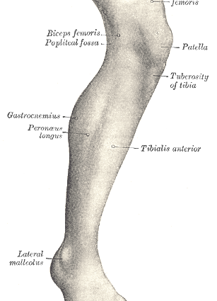
Leg muscles. The leg is made up of three compartments made up of different muscles (1):
- the anterior compartment which is composed of four muscles: the tibialis anterior, the extensor digitorum longus, the extensor hallucis longus and the third fibular
- the lateral compartment which is composed of two muscles: the fibular longus muscle and the fibular short muscle
- the posterior compartment which is made up of seven muscles divided into two groups:
– the superficial compartment which consists of the plantar muscle and the triceps sural muscle, comprising three bundles: the lateral gastrocnemius, the medial gastrocnemius and the solar muscle
– the deep compartment which is made up of the poliphate, the flexor digitorum longus, the flexor hallucis longus and tibialis posterior.
The lateral compartment and the superficial posterior compartment form the calf.
Blood supply to the leg. The anterior compartment is supplied by the anterior tibial vessels, while the posterior compartment is supplied by the posterior tibial vessels as well as the peroneal vessels (1).
Innervation of the leg. The anterior, lateral and posterior compartments are respectively innervated by the deep peroneal nerve, the superficial peroneal nerve and the tibial nerve. (2)
Physiology of the leg
Weight transmission. The leg transfers the weight from the thigh to the ankle (3).
Dynamic sound feeling. The structure and position of the leg contribute to the ability to move and maintain good posture.
Pathologies and pains of the legs
Pain in the legs. The causes of pain in the leg can be varied.
- Bone lesions. Severe pain in the leg may be due to a fracture of the tibia or fibula (or fibula).
- Bone pathologies. Pain in the leg may be due to a bone disease such as osteoporosis.
- Muscular pathologies. The muscles of the legs can be subjected to pain without injury such as cramping or suffer muscle injury such as straining or straining. In the muscles, tendons can also cause pain in the leg, especially during tendinopathies such as tendonitis.
- Vascular pathologies. In case of venous insufficiency in the legs, a feeling of heavy legs may be felt. It is manifested in particular by tingling, tingling and numbness. The causes of heavy leg symptoms are varied. In some cases, other symptoms may appear such as varicose veins due to dilation of the veins or phlebitis due to the formation of blood clots.
- Nerve pathologies. The legs can also be the site of nervous pathologies.
Leg treatments
Drug treatments. Depending on the pathology diagnosed, different drug treatments may be prescribed to reduce pain and inflammation as well as to strengthen bone tissue.
Symptomatic treatment. In the case of vascular pathologies, elastic compression may be prescribed to reduce the dilation of the veins.
Surgical treatment. Depending on the type of pathology diagnosed, surgery may be performed.
Orthopedic treatment. Depending on the type of fracture, the installation of a plaster or a resin can be carried out.
Physical treatment. Physical therapies, through specific exercise programs, can be prescribed such as physiotherapy or physiotherapy.
Leg exams
Physical examination. First, a clinical examination is performed in order to observe and assess the symptoms perceived by the patient.
Medical analysis. In order to identify certain pathologies, blood or urine analyzes can be carried out such as, for example, the dosage of phosphorus or calcium.
Medical imaging examination. X-ray, CT or MRI scintigraphy examinations, or even bone densitometry for bone pathologies, can be used to confirm or deepen the diagnosis.
Doppler ultrasound. This specific ultrasound makes it possible to observe the blood flow.
History and symbolism of the legs
In 2013, The New England Journal of Medicine unveiled an article chronicling the new achievements of bionic prostheses. A team of researchers from the Chicago Rehabilitation Institute has successfully put a robotic leg in place on an amputee patient. The latter is able to control this bionic leg by thought. (4)