


Contents
Cataracts in dogs
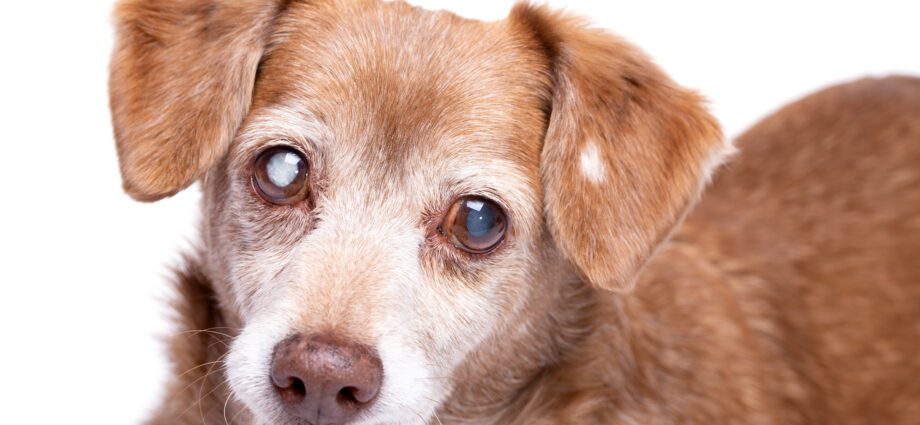
What is cataract in dogs?
The eye is made up of a visible part and an invisible part hidden in the eye socket. In front we find a transparent part called the cornea, with a white part around, the conjunctiva. Behind is the iris which is the diaphragm of the eye then the lens and at the back there is the retina which is a kind of screen in the eye. It is the retina which transmits the nerve message of the image to the brain via the optic nerve. The lens is composed of an outer biconvex capsule and an inner matrix, both are transparent.
The lens is a lens of the eye, it allows light to be focused on the retina. It has a capacity of accommodation which allows it to adapt the vision according to the distance of the object looked at and to keep a clear vision.
Cataracts appear when the proteins in the lens are changed and the matrix becomes completely opaque, preventing light from reaching the retina. The more areas of the lens affected, the more the dog loses his ability to see. When the cataract is advanced the dog totally loses his vision.
Cataracts should not be confused with sclerosis of the lens. You should not be worried about sclerosis of the lens of the eye. As with cataracts, the lens gradually whitens. But this whitening of the lens does not prevent light from passing through and the dog can still see.
What Are the Causes of Cataracts in Dogs?
Cataracts in dogs are very often an age-related disease.
We speak of senile cataract: it preferentially affects dogs over 7 years old. It reaches both eyes and moves slowly.
Another of the main causes is cataract linked to the breed of the dog: it is then a hereditary cataract, so it has a genetic origin. Thus certain breeds of dogs are clearly predisposed to the appearance of cataracts. We can take the example of Yorkshire or the Poodle. This type of cataract being known, we can try to intervene early when it appears to keep the dog’s vision.
Retinal diseases and other causes of inflammation of the eye can cause cataracts to appear in dogs. Thus contusions of the eyeball following shocks or trauma are also causes of the appearance of cataracts in dogs.
When the lens changes position and tilts, we talk about dislocation of the lens. This dislocation is another etiology for cataracts. This dislocation of the lens can occur as a result of inflammation or shock, some breeds like the Shar-Pei are more exposed to dislocation of the lens.
Finally, dogs with diabetes can develop cataracts and lose sight. This diabetic cataract usually develops rapidly and affects both eyes.
Cataract examinations and treatments in dogs
If your dog’s eye and especially your dog’s lens turn white your vet will perform a complete eye exam to determine if there are any underlying causes for the dog’s cataract to appear.
The ophthalmologic examination includes:
- First, an observation from a distance from the eye, we check whether a trauma has not damaged the eyelids or the eye socket, if the eye is not abnormally large (buphthalmos) or protruding (exophthalmos).
- Then if the eye is red and there is conjunctivitis in the dog, corneal tests are carried out.
- In general, if there is a lesion of the lens and in particular if there is a dislocation of the lens, the intraocular pressure (IOP) is measured in order to rule out the suspicion of glaucoma induced by the abnormal displacement of the lens. Glaucoma is an abnormal increase in IOP and poses a risk for loss of the eye. He must be treated urgently if he is present.
- With a view to possible lens surgery to restore sight to the dog, the veterinarian does (or has a veterinarian specializing in ophthalmology) a neurological examination of the retina. In fact, if the retina no longer works or does not transmit images correctly, surgery will be useless and will not restore vision to the dog. This exam is called electroretinography.
The only treatment for canine cataracts is surgery. It is performed by a veterinary ophthalmic microsurgeon and requires very specific equipment, such as an ophthalmic microscope, miniature tools, and an apparatus to lyse and aspirate the lens matrix. For this reason this surgery is very expensive. The veterinarian will make an opening between the cornea and the conjunctiva to introduce his tools, then remove the matrix that has become opaque from inside the lens capsule and replace it with a transparent lens. Finally he makes a microscopic suture of the opening he had made at the start. During the entire surgery, he must hydrate the cornea to prevent it from drying out and inject products to replace the fluids naturally present in the eye and which escape through the surgical opening.
After the surgery you will need to apply a lot of eye drops to your dog’s eye and the ophthalmologist will check the eyes regularly.