


Cancellous bone: all you need to know about this bone in the human body
Deterioration of the internal structure of bone tissue, including cancellous bone, can cause weaker bones and therefore increase complications. But what is a cancellous bone, and what disease can it trigger?
Anatomy of the parasympathetic nervous system
Bones are classified according to their shape and function into five types, each of which meets a specific need: long bones, flat bones, sesamoid (irregular) bone, and short bones.
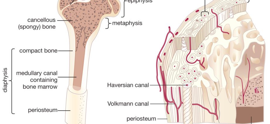
The long bone is made up of an outer layer, the compact bone (smooth and dense) and inside the compact bone is the cancellous bone, which consists of small lamellae or bony trabeculae. The cancellous bone, which is so called because of its terry-like structure, is located at the two ends of the long bone called the epiphyses, on which the greatest compressive forces are exerted.
This type of bone tissue contains the red spinal cord which produces the different blood cells:
- red blood cells;
- white blood cells;
- blood platelets.
In adults, the red pith is found in:
- the bones of the skull;
- The vertebrates ;
- les blades;
- the breastbone;
- the ribs;
- the pelvis;
- the epiphyseal ends of the large long bones.
The amount of red bone marrow decreases with age and is only present in the epiphysis of some bones in adults.
Physiology of cancellous bone
osteoporosis
Type 1 osteoporosis affects the cancellous bone which is mainly due to age since this pathology is linked to aging, to women because they are more exposed due to menopause, and to external factors such as a lack physical activity, calcium and vitamin D deficiency, or excessive alcohol and nicotine consumption.
Osteoclerosis
Characterized by thickening of the cancellous bone resulting in increased bone density.
Abnormalities, pathologies of cancellous bone
osteoporosis
Generalized disease characterized by a decrease in bone mass and bone quality. Osteoporosis can appear when the bone stock is too low or in the event of rapid bone loss.
Osteosclerosis
Characterized by thickening of the cancellous bone resulting in increased bone density.
What treatment for cancellous bone?
osteoporosis
The goal of treatment is to prevent the occurrence of fractures by strengthening the bone tissue.
Before any treatment, the doctor:
- Corrects a potential lack of vitamin D and offers vitamin D supplementation (if necessary) to strengthen bones;
- Ensure adequate calcium intake: dietary intake should be checked and modified if necessary, or prescribe a medication that combines calcium and vitamin D;
- Encourages smoking cessation;
- Encourages the practice of appropriate and sufficient physical activity, which helps to strengthen balance and reduce the risk of falls;
- Ensures the implementation of fall prevention measures.
Then come specific treatments, bisphosphonates, whose molecules “slow down the activity of osteoclasts, the cells that break down bone, thus limiting bone loss” and preventing the risk of fracture.
Osteosclerosis
There is no treatment for osteosclerosis which is regularly irreversible. However, it is possible to consider:
- Taking corticosteroids to strengthen the bones;
- Bone marrow transplantation for osteopetrosis that manifests itself in childhood;
- Plastic surgery to correct severe bone deformities, especially of the face and jaw.
How is the diagnosis made?
osteoporosis
The diagnosis is determined by measuring the density of the bones using densitometry and by x-rays of the dorsolumbar spine which must be performed automatically in order to look for a vertebral fracture that sometimes goes unnoticed and is not painful.
Osteoclerosis
The diagnosis is usually based on clinical signs and a series of x-ray examinations:
- Conventional radiology makes it possible to highlight dense and misshapen bones;
- Computed tomography makes it possible to diagnose possible nerve compressions in the skull;
- Magnetic resonance imaging measures the activity of the bone marrow;
- Bone scintigraphy can identify the densest areas which appear more opaque on the images.