


Contents
calcaneum
The calcaneus (from the Latin calcaneum meaning heel), also called calcaneus, is the largest bone in the tarsus, constituting part of the skeleton of the foot.
Anatomy of the calcaneus
Position. The calcaneus is the largest bone in the tarsus, one of the three parts of the foot skeleton made up of the tarsus, metatarsus, and phalanges (1). The calcaneus is one of the seven bones of the tarsus: the talus, cuboid bone, navicular bone, three cuneiform bones, and calcaneus.
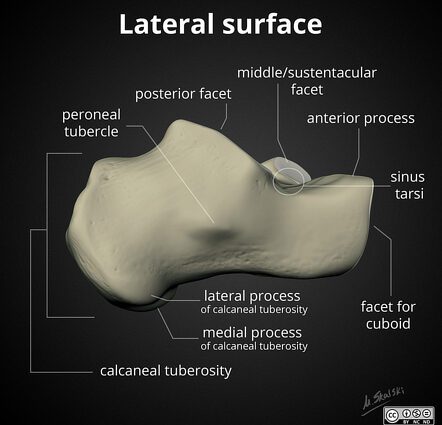
Structure of the calcaneus. The calcaneus is the strongest and largest bone in the foot. The upper surface of the calcaneus articulates with the talus and its anterior surface with the cuboid bone. The calcaneus is made up of:
- sustentaculum tali, a bony projection located on the medial and upper surface, providing support for the talus;
- of the fibular trochea, small crest projecting on the lateral face;
- of the tuberosity of the calcaneus, constituting the protruding posterior surface and forming the heel.
The entire skeleton of the foot, including the calcaneus, is maintained thanks to the numerous ligaments and numerous joints.
Function of the calcaneus
Body weight support. Most of the body’s weight is transmitted from the slope to the ground through the calcaneus (1).
Static and dynamic of the foot. The skeleton of the foot, including the calcaneus, makes it possible in particular to maintain the support of the body and to perform various movements of the foot including the propulsion of the body when walking. (2) (3)
Pathologies of the calcaneus
Foot bone fractures. The skeleton of the foot can be affected by fractures, the most common of which are those of the bones of the metatarsal and calcaneus. (4)
Bone abnormalities. Certain abnormalities can occur in the skeleton of the foot and affect the bones of the metatarsal. These bone abnormalities may in particular be due to malformations, fractures or immobilization. Different cases can be observed: hollow foot, varus foot, flat foot, club foot, or even equine foot. (4)
Maladies of the os. Many diseases can affect the bones and change their structure. Osteoporosis is one of the most common conditions. It constitutes a loss of bone density generally in people over the age of 60. It accentuates bone fragility and promotes bills.
Treatments
Medical treatment. Depending on the disease diagnosed, different treatments may be prescribed to regulate or strengthen bone tissue or reduce pain and inflammation.
Surgical treatment. Depending on the type of fracture, a surgical operation can be performed with the installation of a screw plate, nails or an external fixator.
Orthopedic treatment. Depending on the type of fracture, a plaster cast may be performed.
Examination of the calcaneus
Medical imaging exam. X-ray, CT, MRI, scintigraphy or bone densitometry examinations can be used to assess bone pathologies.
Medical analysis. In order to identify certain pathologies, blood or urine tests can be carried out such as the dosage of phosphorus or calcium.
History
“Little Foot” (in French, petit pied) is the name given to a skeleton ofAustralopithecus prometheusdiscovered in 1994 by paleoanthropologist Ronald J. Clarke. It owes its name “Little Foot” to the small size of the foot bones initially found in a box of bones classified as coming from bovines. After the discovery of these small foot bones, the researchers found 90% of the skeleton: “Little Foot” thus became the most complete Australopithecus skeleton discovered to date. After highly variable dating results, a new method has made it possible to date it to 3,67 million years old (5) (6).